Short Communication - Der Pharma Chemica ( 2023) Volume 15, Issue 3
UPLC method Validation for bioanalysis of Gemcitabine in rat PK study using VAMS methodology (Volumetric Absorptive Microsampling)
Raja Rajeswari K1*, Hafis Jamal Ayyil2 and Kathirvel S22National College of Pharmacy, KMCT Group of Institutions, Manassery, Kozhikode-673602, Kerala, India
Raja Rajeswari K, Department of Pharmaceutical Analysis, Sri Sivani College of Pharmacy, Srikakulam-532 402, Andhra Pradesh, India, Email: drrajarajeswarikatta@gmail.com
Received: 05-May-2023, Manuscript No. dpc-23-97900; Accepted Date: May 31, 2023 ; Editor assigned: 08-May-2023, Pre QC No. dpc-23-97900; Reviewed: 22-May-2023, QC No. dpc-23-97900; Revised: 26-May-2023, Manuscript No. dpc-23-97900; Published: 31-May-2023, DOI: 10.4172/0975-413X.15.3.38-51
Abstract
Volumetric absorptive microsampling (VAMS) is a simple intuitive technique for collecting and quantitative analysis of dried blood samples. It enables the collection of an accurate blood volume regardless of blood hematocrit. A bioanalytical method for the determination of gemcitabine in dried blood supported on VAMS samplers has been validated and used to support a pharmacokinetic study in rat. The calculated PK parameters were comparable to those obtained from blood–water (1:1, v/v) samples. VAMS is demonstrated to be a robust method that simplifies both the blood sample collection and bioanalytical laboratory procedures and generates high quality quantitative data. Waters Acquity UPLC system using an Acquity BEH C18 column (100x2.1 mm, 1.7 μm) was used for chromatographic separation by isocratic elution using acetonitrile-water (35-65) as the mobile phase at a flow rate of 0.5 mL/min. Gemcitabine was administered to rat orally at 3 mg/kg for conducting the PK study and the blood was collected at various time intervals using VAMS sampler which consists of a hydrophilic polymeric tip, absorbs an accurate sample volume within 2–4 s by wicking, attached to a molded plastic handle. The tip is white before use and turns completely red when filled with blood, and the blood samples were processed after collection and analyzed by UPLC. The intra-day and inter-day accuracy of gemcitabine were 93.6–108.7% and 94.2–111.5% respectively, and the precision (RSD, %) was less than 15% for both intra-day and inter-day measurements. Gemcitabine has a good linear relationship in the range of 50-500 ng/mL with r2 value of 0.997. A robust and reliable UPLC method was fully optimized and developed to detect the blood concentration of gemcitabine in rats and the samples were analyzed by Empower software.
Keywords
Gemcitabine; VAMS; UPLC; Validation, Bio analytical; Pharmacokinetics
INTRODUCTION
Gemcitabine is a synthetic pyrimidine nucleoside prodrug, a nucleoside analog in which the hydrogen atoms on the 2' carbon of deoxycytidine are replaced by fluorine atoms [1]. This drug treats cancers including testicular cancer, breast cancer, ovarian cancer, non-small cell lung cancer, pancreatic cancer, and bladder cancer [2]. Gemcitabine is in the nucleoside analog family of medication. It works by blocking the creation of new DNA, which results in cell death [3]. Gemcitabine can increase your risk of bleeding or infection [4]. Even though the drug is approved for medical use in 1995, very less clinical data is available for this and no rapid UPLC method has been reported so far to analyse the bio samples. When gemcitabine is incorporated into DNA it allows a native, or normal, nucleoside base to be added next to it. This leads to "masked chain termination" because gemcitabine is a "faulty" base, but due to its neighbouring native nucleoside it eludes the cell's normal repair system (base-excision repair). Thus, incorporation of gemcitabine into the cell's DNA creates an irreparable error that leads to inhibition of further DNA synthesis, and thereby leading to cell death. The form of gemcitabine with two phosphates attached (dFdCDP) also has activity; it inhibits the enzyme ribonucleotide reductase (RNR), which is needed to create new DNA nucleotides. The lack of nucleotides drives the cell to uptake more of the components it needs to make nucleotides from outside the cell, which also increases uptake of gemcitabine. Gemcitabine is marketed as Gemzar and it is available as intravenous injection. It is approved by the FDA to treat advanced ovarian cancer in combination with carboplatin, metastatic breast cancer in combination with paclitaxel, non-small cell lung cancer in combination with cisplatin, and pancreatic cancer as monotherapy. It is also being investigated in other cancer and tumour types. The aim of the current research is to develop and validate a rapid, reliable, sensitive and simple ultra-performance liquid chromatography method for the quantification of Gemcitabine in whole human blood by Volumetric Absorptive microsampling (VAMS) [5] technique. The advantages of taking microsamples (typically blood samples within the range 10–100 μL), particularly for the determination of rodent pharmacokinetics (PK) and toxicokinetics (TK) has been well documented [6]. The VAMS sampler consists of an absorbent tip, that wicks up an accurate volume of blood (approximately 10 μL), attached to a plastic handle. The volume of blood absorbed is independent of the HCT of the blood. The sample collection procedure involves dipping the tip of the sampler into a pool of blood, for 4–6 s. The sample that is collected is then in the format used for storage and shipping, with only drying and packaging required as additional processing steps. In addition, since the sampling device itself becomes the sample to be analyzed, there is also a reduction in the workflow complexity in the bioanalytical laboratory, with the elimination of the need for aliquotting as with liquid samples, or sub-punching of DBS samples [7, 8]. Further, the design of the sampling device readily enables automation using standard liquid handling robots.
A very few analytical methods are available for the determination of Gemcitabine by chromatographic methods. Although several HPLC [9-16] and LC-MS [17-24] methods are available for bioanalysis but most of them are very expensive and time consuming. Till date there is no UPLC method reported for bioanalysis of Gemcitabine.
The objective of the present work is to develop and validate a simple assay on UPLC (Ultra Performance Liquid Chromatography) using VAMS technique to determine Gemcitabine concentrations in whole human blood. The developed bioassay is validated using internationally accepted criteria. After complete validation, the method was applied to analyze study sample analysis in rats by giving a single oral dose at 3 mg/kg body weight. Data generated from dried VAMS samples is compared to that from VAMS samples extracted before drying and that from the more conventional approach of blood sampling, where whole blood is quantitatively diluted with water. In addition, the effect of HCT, storage and initial blood temperature are investigated.
EXPERIMENTAL
Instrumentation and Chromatographic Conditions (2)
UPLC–UV Analysis (2a)
The LC system consisted of a Waters Acquity UPLC with Empower software equipped with a photodiode array detector. A Acquity BEH C18 column (100x2.1 mm, 1.7 μm particle size) from Waters was used as stationary phase and temperature maintained at 20°C. The mobile phase consisted of Acetonitrile and water (35:65) in isocratic mode pumped at a flow rate of 0.5 mL/min. Analysis was performed for 5 min at the detection wavelength of 275 nm and the injection volume was 5 μL. The autosampler maintained at 4°C.
Chemicals (2b)
Gemcitabine and internal standard (2’-deoxycytidine) are purchased from Sigma–Aldrich Trading Co., Ltd. (Shanghai, China). Acetonitrile and methanol of HPLC grade and all other chemicals were obtained from Merck (Mumbai, India). Water used in the entire analysis was prepared from Milli-Q water purification system from Millipore (Milford, MA, USA). Biological matrix (whole human blood) was obtained from Vimta Labs (Hyderabad, India) and stored at −20°C until use.
Preparation of Calibrators and QC Samples (2c)
A standard stock solution of Gemcitabine was prepared by dissolving standard 50 mg of Gemcitabine into 50 ml volumetric flask, to this added 30 ml of methanol and sonicated for 10 minutes at a temperature not exceeding 20°C. Allowed the solution to attain room temperature and then diluted to the volume with methanol to have a solution with a concentration of 1000 μg/mL. Calibration standard and quality control (QC) samples were prepared by adding corresponding working solutions with drug-free human blood. A volume of 10 mL of appropriate diluted stock solution at different concentrations and 10 mL of IS at a fixed concentration were spiked into 200 μL of human blood to yield final concentrations of calibration samples 50, 100, 150, 200, 250, 300, 400 and 500 ng/mL. The final concentration of IS was 100 ng/mL. Similarly, QC samples were prepared at four concentration levels LLOQ (50 ng/mL), LQC (150 ng/mL) MQC (250 ng/mL) and HQC (400 ng/mL) in a similar manner to the calibration standards but from an independent stock solution.
Sample preparation (2d)
Analytes were extracted from blood by employing VAMS method, vortexes for 1min and then centrifuged at 10,000 rotations per minute for 10 min on refrigerated centrifuge at 4°C. The supernatant layer was separated and filtered through 0.45 μm syringe filters and 10 μL of the solution was injected for UPLC analysis.
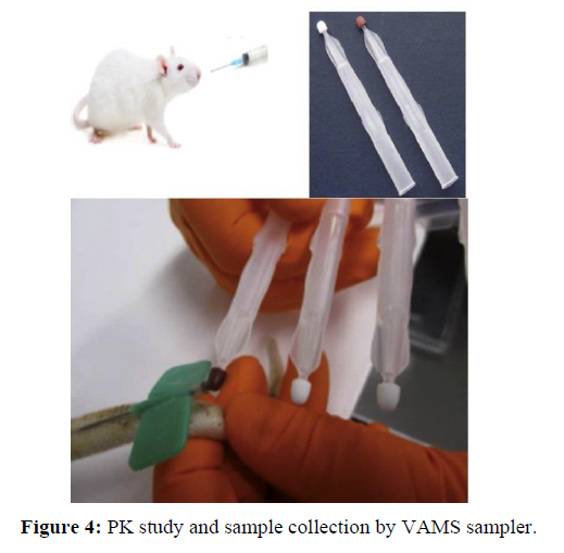
The newer sampling technique, Volumetric Absorptive Micro sampling (VAMS) allows reduction of volume from milliliter to microliter (sample volume ∼ 10μl). The micro sampling devices (Mitra®) have overcome almost all drawbacks of conventional sampling with a few additional benefits. A novel dried blood sampler, VAMS, allows consistent blood volume regardless of Hematocrit (Hct). It is available in a configuration of 2 samples with volume 10, 20 and 30 μl. A sampler of 10 and 20 μl is usually used for sampling in animals and 30 μl in humans. The unique device consists of an absorbent polymeric tip which enables the collection of fixed, a small volume of blood by capillary action. The sample is obtained either by finger or heel prick for humans and tail vein in rodents. During collection, the sampler is filled by holding the handle at an angle of 45° and dipping only the tip into blood drop and allowing it to fill. The tip of the sampler should not be completely plunged into the blood sampler. This may cause overfilling of the sample. The device is self-indicating i.e. when the tip is filled, it turns red. The tip is attached to a handle, which is designed in a way that prevents the sampler tip coming into contact with surfaces during storage and shipping. Samples can be shipped or stored at room temperature. VAMS device ensures the homogeneity of the sample, as a precise volume is absorbed on to the tip. During sample preparation, either the tip is removed from the handler or the whole device is used. This device enables ease of sample pretreatment as the centrifugation step of the liquid matrix and sub-punching step of DBS (Dried blood spot) is subtracted. Moreover, the sampler is configured to fit in manual or automated extraction devices. The greatest advantage of VAMS over DBS is that VAMS enables the precise and accurate collection of blood volumes for quantitative bioanalysis. The dried VAMS calibration and QC samples were extracted by removing the tip from its sampler by pulling the tip against the inside of the extraction tube, to which 200 μL of methanol containing internal standard was added. The sealed tubes were mixed on a lateral shaker for an hour. The extracts were diluted 9-fold with methanol–water (1:1, v/v), prior to analysis for gemcitabine by UPLC.
Preparation and extraction of wet samples from VAMS samplers (2e)
In order to prepare wet VAMS samples, blood was absorbed onto the VAMS tip as previously described, and then immediately removed from the holder by pulling the tip against the side of a 1.4 mL Micronics tube to which water (100 μL) had been added. After sealing, the tube was vortex mixed and allowed to stand for 1 h to allow cell lyses to occur. The wet VAMS blood–water samples were either used immediately, or stored frozen at -20◦C. Gemcitabine was extracted from aliquots of the wet VAMS blood–water samples by protein precipitation, following the addition of 5 volumes of methanol containing internal standard (5 μg/mL) and EDTA, followed by centrifugation at 5000 rpm at 4C for 10 min. The supernatant was diluted 2-fold with methanol-water (1:1, v/v) prior to analysis by UPLC.
Preparation and extraction of blood–water (1:1, v/v) samples (2f)
Blood–water samples were prepared by mixing equal volumes of blood and water (100 μL of each) and allowing them to stand for an hour. These were either used immediately, or stored frozen at -20C. The extraction procedure for blood–water samples was the same as for wet VAMS samples, except 10 volumes of methanol containing internal standard was used at the precipitation stage and the supernatant was diluted 9-fold with methanol-water (1:1, v/v) prior to analysis by UPLC.
ANALYTICAL VALIDATION
All validation experiments were performed according to the Bio analytical Method Validation Guidance for Industry [25] and the ICH guidelines [26] on validation of bio-analytical methods.
Assay Specificity and Selectivity (3a)
Specificity was assessed by verifying the absence of significant interference in the biological control medium with regard to the retention time of the compound (s) to be assayed. The specificity of the method was confirmed by comparing chromatograms of blank matrix, spiked matrix with analyte at LOQ concentration. No interfering endogenous peaks were observed around the retention time.
Linearity (3b)
A calibration curve was prepared within the range of 50 to 500 ng/mL gemcitabine in each run. Half of the calibration samples were analyzed at the beginning of the run and half at the end. The simplest calibration model and weighting procedure were used. The calculations of the curve’s parameters were based on the ratio of the peak areas of gemcitabine/IS versus the concentration of gemcitabine. Gemcitabine concentrations for samples were calculated from the curve’s equation obtained by means of linear regression.
Accuracy of back-calculated calibration samples should be within ±15% of the corresponding nominal concentration, except at the lowest concentration level, where the accuracy should be within ±20%. Per calibration curve, a maximum of 33% of the calibration samples, except the LLOQ and upper limit of quantification (ULOQ, 500 ng/mL), may differ from these specifications. At least 6 concentration levels were represented in each curve.
Matrix Effect, Extraction Recovery, and Process Efficiency (3c)
The influence of the matrix on the quantification of Gemcitabine was monitored using a comparison of: (1) the instrument response for the low, medium, and high QCs (n = 4 per level) injected directly in mobile phase (neat solutions), (2) the same amount of analyte added to extracted blank samples (post extraction spiked samples), and (3) the same amount of analyte added to the biological matrix before extraction (pre extraction spiked samples). Total process efficiency was calculated from the ratio of mean peak areas of Gemcitabine in extracted validation samples versus neat unextracted samples. This term accounts for any loss in signal attributable to the extraction process or matrix effect. Extraction recovery was calculated from the ratio of mean peak areas of Gemcitabine in extracted validation samples versus blank samples spiked after extraction. The absolute matrix effect was calculated from the ratio of mean peak areas of Gemcitabine in blank samples spiked after extraction versus neat unextracted samples. If the ratio was 85% or 115%, an exogenous matrix effect was inferred.
Matrix Variability (3d)
To confirm that the biological matrix would not interfere with the assay, the selectivity of the developed method was tested by analyzing 6 different lots of blank blood samples and also 6 different lots of blank urine samples spiked with IS at the LLOQ level (n = 3 per lot), and blank blood samples with no IS (n = 3 per lot) against a calibration curve. The results for the LLOQ samples were considered acceptable if the precision from each matrix lot was ±20% and the accuracy was within the range of 80%–120%. The acceptance criterion for the analysis of the blank samples from the 6 individual lots was based on the raw peak areas found at the retention times of Gemcitabine and IS. No more than 10% of the blank samples could have peak areas greater than 20% of the average peak area of Gemcitabine in the LLOQ QCs.
Stability studies (3e)
Stability evaluations were performed in both aqueous and matrix based samples. Stability evaluations in matrix were performed against freshly spiked calibration standards using freshly prepared quality control samples (comparison samples). Gemcitabine stability in blood was evaluated by performing bench top stability, long-term stability, short term stability and freeze-thaw stability. The processed samples were studied for stability in auto sampler at 10°C. Stability in blood was evaluated at both low and high QC level by comparing the mean response ratio of stability samples against the comparison samples.
RESULTS AND DISCUSSION
Chromatographic and detection parameters (4a)
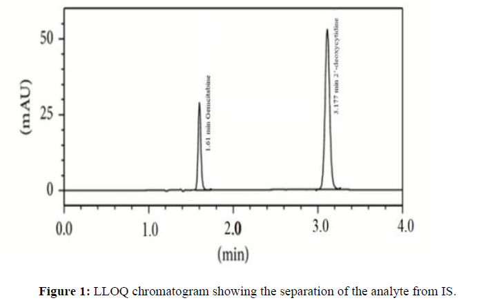
Optimal chromatographic conditions were obtained after running different mobile phases with a reversed-phase C18 column. The different columns tried were Symmetry C18, Luna C18 and Zorbax C18. The best results were observed with the Acquity UPLC BEH C18 column (2.1 mm × 100 mm, 1.7 μm particle size) using acetonitrile and water (35:65) as mobile phase. Variation of the column temperature between 20 and 30°C did not cause significant change in the resolution, however changes in retention time were observed. The column was used at 20°C at a flow rate of 0.5 mL/min. The method allowed the separation of analyte with IS in 5 min (Figure 1) runtime.
Specificity, Linearity, Accuracy and Precision (4b)
The specificity of method was confirmed by comparing chromatograms of blank matrix, spiked matrix with analyte at LOQ concentration. No interfering endogenous peaks were observed around their retention times. The eight point calibration curve for the analyte showed a linear correlation between concentration and peak area. Calibration data (Table 1) indicated the linearity (r2 > 0.99) of the detector response for all standard solutions from 50 to 500 ng/mL The limits of detection by UPLC was found to be 20 ng/mL and LOQ was found to be 50 ng/mL. All standards and samples were injected in triplicate.
| Concn (ng/mL) | Peak Area |
|---|---|
| 50 | 1835 |
| 100 | 3525 |
| 150 | 5355 |
| 200 | 7368 |
| 250 | 9340 |
| 300 | 10951 |
| 400 | 14402 |
| 500 | 19309 |
| y = 38.141x - 286.23 | |
| R² = 0.997 | |
Multiple injections showed that the results are highly reproducible and showed low standard error. A recovery experiment was performed to confirm the accuracy of the method. Blank blood was spiked with Low QC, Mid QC and High QC levels of the standard stock solution and then extracted and analysed under optimized conditions. The extraction recoveries of all samples from human blood were in the range of 93.7-112.4% with relative standard deviations less than 10.0%, which indicates the sample preparation technique is suitable for extracting (Table 2).
| LLOQ QC | LOW QC | MID QC | HIGH QC | |||||
|---|---|---|---|---|---|---|---|---|
| 50 ng/mL | 150 ng/mL | 250 ng/mL | 400 ng/mL | |||||
| Conc found | % Recovery |
Conc found | % Recovery |
Conc found | % Recovery |
Conc found | % Recovery |
|
| Recovery | 56.204 | 112.407 | 156.722 | 104.466 | 250.114 | 99.927 | 394.569 | 98.642 |
| 49.908 | 99.816 | 149.448 | 99.617 | 251.196 | 100.359 | 400.841 | 100.21 | |
| 55.226 | 110.451 | 140.671 | 93.767 | 253.271 | 101.188 | 393.941 | 98.485 | |
| 50.601 | 101.202 | 162.704 | 108.454 | 250.863 | 100.226 | 396.766 | 99.191 | |
| 55.107 | 110.214 | 156.716 | 104.462 | 253.486 | 101.274 | 393.556 | 98.389 | |
| 52.204 | 104.408 | 142.322 | 94.868 | 256.993 | 102.675 | 408.985 | 102.246 | |
| N | 6 | 6 | 6 | 6 | 6 | 6 | 6 | 6 |
| Mean | 53.208 | 106.416 | 151.431 | 100.939 | 252.654 | 100.942 | 398.11 | 99.527 |
| SD | 2.659 | 8.783 | 2.517 | 5.97 | ||||
| CV(%) | 4.997 | 5.8 | 0.996 | 1.499 | ||||
Intra-day and inter-day precision of the method was determined by analysing QC samples on two consecutive days and the obtained intra-day accuracies were in the range of 93.6–108.7% and inter-day accuracies were in the range of 94.2–111.5%. The recovery results are displayed in (Table 3) and (Table 4).
| Gemcitabine | ||||||||
|---|---|---|---|---|---|---|---|---|
| LLOQ QC | LOW QC | MID QC | HIGH QC | |||||
| 50 ng/mL | 150 ng/mL | 250 ng/mL | 400 ng/mL | |||||
| Intra- day | Concn found | % Recovery |
Concn found | % Recovery |
Concn found | % Recovery |
Concn found | % Recovery |
| 52.914 | 105.827 | 149.646 | 99.618 | 244.79 | 97.684 | 387.033 | 96.758 | |
| 54.313 | 108.626 | 140.639 | 93.622 | 251.236 | 100.257 | 397.367 | 99.342 | |
| 51.447 | 102.894 | 143.223 | 95.342 | 246.306 | 98.289 | 395.875 | 98.969 | |
| 54.345 | 108.69 | 144.673 | 96.307 | 250.196 | 99.842 | 403.859 | 100.965 | |
| 52.886 | 105.772 | 145.144 | 96.621 | 255.884 | 102.111 | 384.929 | 96.232 | |
| 52.779 | 105.559 | 148.89 | 99.114 | 252.78 | 100.873 | 397.899 | 99.475 | |
| N | 6 | 6 | 6 | 6 | 6 | 6 | 6 | 6 |
| Mean | 53.114 | 106.228 | 145.369 | 96.771 | 250.199 | 99.843 | 394.494 | 98.623 |
| SD | 1.09 | 3.412 | 4.11 | 7.164 | ||||
| CV(%) | 2.051 | 2.347 | 1.643 | 1.816 | ||||
| Gemcitabine | ||||||||
|---|---|---|---|---|---|---|---|---|
| LLOQ QC | LOW QC | MID QC | HIGH QC | |||||
| 50 ng/mL | 150 ng/mL | 250 ng/mL | 400 ng/mL | |||||
| Concn found | % Recovery |
Concn found | % Recovery |
Concn found | % Recovery |
Concn found | % Recovery |
|
| Inter-day | 55.496 | 110.991 | 152.974 | 101.968 | 252.305 | 100.803 | 410.627 | 101.922 |
| 55.473 | 110.947 | 141.284 | 94.176 | 254.237 | 101.574 | 404.305 | 100.353 | |
| 55.771 | 111.542 | 154.625 | 103.068 | 252.745 | 100.978 | 402.002 | 99.781 | |
| 55.193 | 110.386 | 148.136 | 98.743 | 251.368 | 100.428 | 406.329 | 100.855 | |
| 55.02 | 110.039 | 160.512 | 106.992 | 251.45 | 100.461 | 399.015 | 99.039 | |
| 49.231 | 98.462 | 145.385 | 96.909 | 256.47 | 102.466 | 413.311 | 102.588 | |
| N | 6 | 6 | 6 | 6 | 6 | 6 | 6 | 6 |
| Mean | 54.364 | 108.728 | 150.486 | 100.309 | 253.096 | 101.118 | 405.931 | 100.756 |
| SD | 2.528 | 6.929 | 1.956 | 5.34 | ||||
| CV(%) | 4.65 | 4.604 | 0.773 | 1.316 | ||||
To investigate carry-over from one sample to the other in the auto sampler, each validation run containing a calibration curve included a blank sample analysed directly after the sample at the ULOQ calibration level. The response of interfering peak (s) in the blank sample should not exceed 20% of the response of the component peak at the LLOQ calibration sample concentration. To demonstrate that the method is suitable for blood sample with test compound concentration higher than the ULOQ, the dilution integrity was assessed using validation samples spiked with the test compound at 2-, 4-, and 10-fold the concentration of the high QC. The dilution test was performed by increasing the concentration of IS by the appropriate dilution factor. After extraction, the dry extract was taken up with a volume of injection solvent also multiplied by the same factor. Accuracy of the calculated concentrations within the range of 85%–115% of the nominal values would suggest that samples containing Gemcitabine at a higher concentration than the ULOQ can be diluted using the above tested dilution method.
Stability evaluations were performed in both aqueous and matrix-based samples. The stock solutions were stable for a period of 24 h at room temperature and for 60 days at 1–10°C. Stability evaluations in matrix were performed against freshly spiked calibration standards using freshly prepared quality control samples (comparison samples). The processed samples were stable up to 36 h in auto sampler at 10°C. The long-term matrix stability was evaluated at −20°C over a period of 60 days. No significant degradation of analytes was observed over the stability duration and conditions. The long-term stability results presented in (Table 5) were within 85–115%. Stability in blood was evaluated at both low and high QC level by comparing the mean response ratio of stability samples against the comparison samples. The short-term stability of analyte at room temperature was within 85–115% up to 24 hours.The stability results presented in (Table 6) and (Table 7)
| Long term stability after 60 days | Gemcitabine | |||
|---|---|---|---|---|
| 0 Hr-Low QC |
0 Hr- HQC |
Day-60- LQC |
Day-60- HQC |
|
| Conc found | Conc found |
Conc found |
Conc found | |
| 157.217 | 386.091 | 152.355 | 385.579 | |
| 150.549 | 404.247 | 153.69 | 399.456 | |
| 151.185 | 408.218 | 151.05 | 411.678 | |
| 149.622 | 392.919 | 147.75 | 381.747 | |
| 142.374 | 406.491 | 145.35 | 389.795 | |
| 152.96 | 419.751 | 146.67 | 417.106 | |
| N | 6 | 6 | 6 | 6 |
| Mean | 150.651 | 402.953 | 149.478 | 397.56 |
| SD | 4.864 | 11.909 | 3.359 | 14.414 |
| CV(%) | 3.229 | 2.955 | 2.247 | 3.626 |
| % Change |
n/a | n/a | -0.779 | -1.338 |
| Short term stability | Gemcitabine | |||||
|---|---|---|---|---|---|---|
| LOW QC | ||||||
| 150 ng/mL | ||||||
| 0 Hour | 4 Hour | 24 Hour | ||||
| Conc found |
% Recovery |
Conc found |
% Recovery |
Conc found |
% Recovery |
|
| 157.217 | 104.812 | 152.55 | 101.7 | 155.88 | 103.92 | |
| 150.549 | 100.366 | 153.015 | 102.01 | 149.91 | 99.94 | |
| 151.185 | 100.79 | 147.18 | 98.12 | 155.811 | 103.874 | |
| 149.622 | 99.748 | 144.834 | 96.556 | 158.746 | 105.83 | |
| 142.374 | 94.916 | 143.442 | 95.628 | 139.535 | 93.023 | |
| 152.96 | 101.973 | 145.594 | 97.062 | 155.769 | 103.846 | |
| N | 6 | 6 | 6 | 6 | 6 | 6 |
| Mean | 150.651 | 100.434 | 147.769 | 98.513 | 152.608 | 101.739 |
| SD | 4.864 | 4.069 | 7.026 | |||
| CV(%) | 3.229 | 2.754 | 4.604 | |||
| % Change |
n/a | -1.913 | 1.299 | |||
| Short term stability | Gemcitabine | |||||
|---|---|---|---|---|---|---|
| High QC | ||||||
| 400 ng/mL | ||||||
| 0 Hour | 4 Hour | 24 Hour | ||||
| Conc found |
% Recovery |
Conc found |
% Recovery |
Conc found |
% Recovery |
|
| 386.091 | 96.523 | 392.287 | 98.072 | 381.136 | 95.284 | |
| 404.247 | 101.062 | 400.885 | 100.221 | 394.437 | 98.609 | |
| 408.218 | 102.055 | 411.518 | 102.879 | 400.426 | 100.106 | |
| 392.919 | 98.23 | 395.315 | 98.829 | 388.471 | 97.118 | |
| 406.491 | 101.623 | 412.094 | 103.023 | 409.955 | 102.489 | |
| 419.751 | 104.938 | 413.865 | 103.466 | 406.721 | 101.68 | |
| N | 6 | 6 | 6 | 6 | 6 | 6 |
| Mean | 402.953 | 100.738 | 404.327 | 101.082 | 396.858 | 99.214 |
| SD | 11.909 | 9.392 | 11 | |||
| CV(%) | 2.955 | 2.323 | 2.772 | |||
| % Change |
n/a | 0.341 | -1.513 | |||
Gemcitabine was stable up to 10 h on bench top at room temperature and over 3 freeze–thaw cycles. In human blood, the freeze-thaw study was carried out and the results are presented in (Tables 8 and 9).
| Freeze Thaw Cycle- III | Gemcitabine | |||
|---|---|---|---|---|
| Freeze Thaw Cycle-III below -20°C | ||||
| LOW QC | HIGH QC | |||
| 150 ng/mL | 400 ng/mL | |||
| Conc found | % Recovery | Conc found | % Recovery | |
| 143.67 | 95.78 | 391.251 | 97.813 | |
| 148.89 | 99.26 | 384.471 | 96.118 | |
| 145.542 | 97.028 | 398.997 | 99.749 | |
| 143.367 | 95.578 | 396.982 | 99.245 | |
| 157.107 | 104.738 | 393.286 | 98.322 | |
| 159.63 | 106.42 | 400.974 | 100.243 | |
| N | 6 | 6 | 6 | 6 |
| Mean | 149.701 | 99.801 | 394.327 | 98.582 |
| SD | 7.041 | 6.012 | ||
| CV(%) | 4.703 | 1.525 | ||
| Freeze Thaw Cycle-III | Gemcitabine | |||
|---|---|---|---|---|
| Freeze Thaw Cycle-III below -50°C | ||||
| LOW QC | HIGH QC | |||
| 150 ng/mL | 400 ng/mL | |||
| Conc found | % Recovery | Conc found | % Recovery | |
| 143.13 | 95.42 | 408.431 | 102.108 | |
| 147.39 | 98.26 | 399.229 | 99.807 | |
| 150.6 | 100.4 | 387.387 | 96.847 | |
| 147 | 98 | 386.616 | 96.654 | |
| 138.39 | 92.26 | 387.888 | 96.972 | |
| 144.51 | 96.34 | 411.111 | 102.778 | |
| N | 6 | 6 | 6 | 6 |
| Mean | 145.17 | 96.78 | 396.777 | 99.194 |
| SD | 4.203 | 11.115 | ||
| CV(%) | 2.895 | 2.801 | ||
The variability of the matrix effect in whole human blood has resulted a very minute change in the recovery of middle concentration of calibration curve. The results of Matrix effect area presented in (Table 10).
| Gemcitabine | ||
|---|---|---|
| Extracted | ||
| Unit No. | Neat standard sample |
blank plus spiked |
| Concentration | sample peak | |
| concentration | ||
| Unit No.: 1 | 9818 | 9040 |
| Unit No.: 2 | 9194 | 8960 |
| Unit No.:3 | 9391 | 9010 |
| Unit No.: 4 | 9038 | 8828 |
| Unit No.: 5 | 9789 | 9677 |
| Unit No.: 6 | 9482 | 9274 |
| N | 6 | 6 |
| Mean | 9452 | 9131.5 |
| SD | 313.083 | 304.15 |
| CV(%) | 3.312 | 3.331 |
| Matrix effect (%) |
0.966 | |
Auto-sampler Carry-Over Test (4c)
To investigate carry-over from one sample to the other in the auto sampler, each validation run containing a calibration curve included a blank sample analysed directly after the sample at the ULOQ calibration level. The response of interfering peak (s) in the blank sample should not exceed 20% of the response of the component peak at the LLOQ calibration sample concentration.
Dilution Integrity Test (4d)
To demonstrate that the method is suitable for a blood sample with test compound concentration higher than the ULOQ, the dilution integrity was assessed using validation samples spiked with the test compound at 2-, 4-, and 10-fold the concentration of the high QC. The dilution test using blood samples was performed by increasing the concentration of IS by the appropriate dilution factor. After extraction, the dry extract was taken up with a volume of injection solvent also multiplied by the same factor. Accuracy of the calculated concentrations within the range of 85%–115% of the nominal values would suggest that a blood sample containing Gemcitabine at a higher concentration than the ULOQ can be diluted using the above tested dilution method.
Effect of blood temperature (4e)
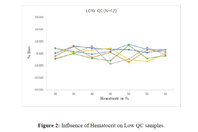
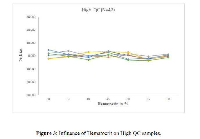
The ruggedness of the assay to variations in the temperature of the blood used to prepare VAMS samples was assessed by comparing the bias of dried VAMS samples generated at low and high QC levels from pools of blood held at 4◦C, ambient temperature (25°C) and 37°C. The maximum bias observed, against a calibration line prepared at ambient temperature, was 11% and the maximum with-in run precision was 5.8% indicating that the temperature of the blood used to generate the samples did not influence the observed concentration. The effect of Hematocrit on the volume of blood absorbed was investigated on low QC (Figure 2) and high QC level (Figure 3) and proved to be promising over an acceptable range.
Application of the method to pharmacokinetic study in Rat (4f)
Wistar rats (220±20 g) used were maintained in a clean room at a temperature between 22±2°C with 12 h light/dark cycles and a relative humidity rate of 50±5%. Rats were housed in cages with a supply of normal laboratory feed with water ad libitum. For all of the studies, the animals (n=6) were deprived of food 12 h before dosing, but had free access to water. In order to verify the sensitivity and selectivity of the developed method in a real-time situation, the developed UPLC method was successfully applied to a pharmacokinetic study by administration of Gemcitabine as single solution to six male wistar rats by oral route using BD syringe attached with oral gavage needle (size 18) at the dose of 3 mg/kg body weight (Figure 4). Approximately, a few drops of blood, drawn by dipping the tips of VAMS samplers into the blood in such a way that the tip just broke the liquid surface. The tips took between 2 and 4 s to completely absorb the blood and fill with color, depending upon the HCT of the blood and the depth to which they were immersed. Although the tip was considered full when it had completely colored, it was held for an additional 2 s in the blood pool before being removed and dried. Care was taken during the filling process to ensure that tips were not submerged past the shoulder. The VAMS samples were dried for a minimum of two hours, in freely circulating laboratory air (21°C, 55% relative humidity, controlled but not monitored) in such a way that the tips did not touch each other or their surroundings. The VAMS samples were extracted by removing the tip from its sampler by pulling the tip against the inside of the extraction tube, to which 200 μL of methanol containing internal standard (100 ng/mL) was added. The sealed tubes were mixed on a lateral shaker for an hour. The extracts were diluted with methanol–water and centrifuged in diluent at 10,000 rpm for 10 min. The obtained supernatant samples were transferred into pre-labeled micro vials. The time intervals for the sample collection were 0 (predose), 0.5, 1, 2, 4, 6, 8, 12, 24 and 48 h (postdose).
The blood samples thus obtained were stored at –30°C till analysis. Post analysis the pharmacokinetic parameters were computed using WinNonlin® software version 5.2 and SAS® software version 9.2.
The pharmacokinetic parameters evaluated were Cmax (maximum observed drug concentration during the study), AUC0-48(area under the blood concentration–time curve measured 48 hours, using the trapezoidal rule), Tmax (time to observe maximum drug concentration), Kel (apparent first order terminal rate constant calculated from a semi-log plot of the blood concentration versus time curve, using the method of least square regression) and t1/2 (terminal half-life as determined by quotient 0.693/Kel).
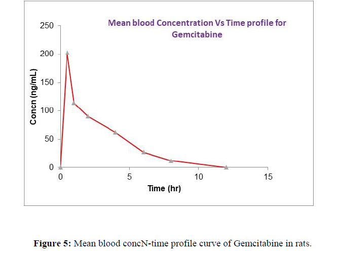
All the samples were analyzed by the developed method and the mean concentrations vs time profile of Gemcitabine is shown in (Figure 5). The pharmacokinetic parameters estimated are shown in (Table 11).
| Parameter | Gemcitabine |
|---|---|
| Cmax (ng/mL) | 202.653 ± 20.551 |
| Tmax (h) | 0.5 ± 0.025 |
| t1/2 (h) | 0.5 ± 0.222 |
| Kel (h-1) | 0.0693 ± 0.046 |
Incurred sample reanalysis (ISR) (4g) Re-analysis of all the dried VAMS, wet VAMS and blood–water (1:1, v/v) study sample sets demonstrated satisfactory ISR results between the original and the repeat result being within 20% of the mean of the two values. The lower agreement rate for the dried VAMS compared to the other two groups probably reflects the fact that the original and repeat dried samples were derived from physically separate sampling events with the VAMS device. Actually, the assay original and repeat analyses for the wet VAMS and blood–water samples were derived from the same liquid pool after the addition of water.
CONCLUSION
Apart from the UPLC method validation, it has also been demonstrated that the changes in assay bias and analyte recovery with HCT are acceptable with VAMS device. It was also demonstrated that temperature of the blood did not affect the assay result obtained. Thus, VAMS tips can be filled from blood straight from the rat tail with a suitable blood draw technique, without having to wait for it to equilibrate to an ambient temperature. One of the rationales for adopting a micro sampling approach is to reduce the amount of blood drawn at each sampling time point, which includes not just the blood collected for the analysis, but also any spilt blood and losses that occurs during staunching of the wound. Although the VAMS tips were overwhelmed, there is good agreement between the replicate VAMS samples for both dry and wet samples, taken at the same time point. The concentrations between the original and replicate results obtained for the dry VAMS samples showed 10% of the samples having a difference greater than 20%. This comparison complies with the ISR criteria and indicating that the volume of blood collected on the tip at any one time point was consistent. Thus the VAMS technique has the ability to replace DBS for quantitative bio analysis, since it retains all the recognized advantages of DBS as well as making the sample collection process simpler, and reduce the work flow within the bio-analytical laboratory and minimizes the effect of HCT on assay bias.
ACKNOWLEDGMENT
Statements and Declarations
Funding (5a)
The authors declare that no funds, grants, or other support were received during the preparation of this manuscript.
Conflict of Interest (5b)
The authors have no relevant financial or non-financial interests to disclose.
Author Contributions (5c)
All authors contributed to the study conception and design. All authors read and approved the final manuscript.
REFERENCES
- Brown K, Weymouth-Wilson A, Linclau B. Carbohydrate Research .2015, 406: p. 71–5.
- National Cancer Institute (2006-10-05). FDA Approval for Gemcitabine Hydrochloride. National Cancer Institute.
- Abbruzzese JL, Grunewald R, Weeks EA, et al., Journal of Clinical Oncology.1991, 9: p. 491–498.
- Drugs.com
- Londhe V, Rajadhyaksha M. J Pharm Biomed Anal. 2020, 182: p. 113102.
- Spooner N. Bioanalysis.2013, 5:p. 879–883.
- Denniff P, Spooner N. Anal. Chem. 2014, 86:p. 8489–8495.
- Spooner N, Denniff P, Michaelsen L, et al., Bioanalysis .2014.
- Freeman KB, Anliker S, Hamilton M, et al., Journal of Chromatography B: Biomedical Applications .1995, 665:p. 71–181.
- Xu H, Paxton J, Lim J, et al., Journal of Pharmaceutical and Biomedical Analysis. 2014, 98:p. 371–378.
- Lanz C, Früh M, Thormann W, et al., Journal of Separation Science .2007,30:p. 1811–1820.
- Losa R, Sierra MI, Gión MO et al. Journal of Chromatography B: Analytical Technologies in the Biomedical and Life Sciences. 2006, 840:p. 44–49.
- Yilmaz B, Kadıoglu Y. Il-Farmaco. 2004, 59:p. 425–429.
- Keith B, Xu Y, Grem JL. Journal of Chromatography B .2003, 785: p. 65–72.
- Yilmaz B, Kadıoğlu Y, Aksoy Y, Journal of Chromatography B: Analytical Technologies in the Biomedical and Life Sciences. 2003, 791:
- Xu Y, Keith B, Grem JL, Journal of Chromatography B. 2004, 802:p. 263–270.
- Bowen C, Wang S, Licea-Perez H, Journal of Chromatography B: Analytical Technologies in the Biomedicaland Life Sciences.2009, 877:p. 2123–2129.
- Kirstein MN, Hassan I, Guire D, et al., Journal of Chromatography B: Analytical Technologies in the Biomedical and Life Sciences. 2006, 835:p. 136–142.
- Little JL, Wempe MF, Buchanan CM, Journal of Chromatography B: Analytical Technologies in the Biomedical and Life Sciences. 2006, 833:p. 219–230.
- Liu Y, Zhang W, Yang Y. Talanta. 2008, 77:p. 412–421.
- Marangon E, Sala F, Caffo O, et al., Journal of Mass Spectrometry. 2008, 43:p. 216–223.
- Pisano R, Breda M, Grassi S, et al., Journal of Pharmaceutical and Biomedical Analysis. 2005, 38:p. 738–745.
- Pucci V, Di Palma S, Alfieri A, et al., Journal of Pharmaceutical and Biomedical Analysis. 2009, 50:p. 867–871.
- Wang LZ, Goh BC, Lee HS, et al., Therapeutic Drug Monitoring. 2003, 25:p. 552–557.
- Food and Drug Administration. Guidance for Industry: Bioanalytical Method Validation Center for Drug Evaluation and Research. Silver Springs.
- ICH guideline M10 on bio analytical method validation and study sample analysis.
Indexed at, Google Scholar, Crossref
Indexed at, Google Scholar, Crossref
Indexed at, Google Scholar, Crossref
Indexed at, Google Scholar, Crossref
Indexed at, Google Scholar, Crossref
Indexed at, Google Scholar, Crossref
Indexed at, Google Scholar, Crossref
Indexed at, Google Scholar, Crossref
Indexed at, Google Scholar, Crossref
Indexed at, Google Scholar, Crossref
Indexed at, Google Scholar, Crossref
Indexed at, Google Scholar, Crossref
p. 103–109.
Indexed at, Google Scholar, Crossref
Indexed at, Google Scholar, Crossref
Indexed at, Google Scholar, Crossref
Indexed at, Google Scholar, Crossref
Indexed at, Google Scholar, Crossref
Indexed at, Google Scholar, Crossref
Indexed at, Google Scholar, Crossref
Indexed at, Google Scholar, Crossref
Indexed at, Google Scholar, Crossref
Indexed at, Google Scholar, Crossref

