Review Article - Der Pharma Chemica ( 2024) Volume 16, Issue 2
The Effects of Bioactive Components Derived from Fruit on Different Types of Cancer: Mechanistic Pathways and Settings Associated to Stress
Sandhiya*Sandhiya, Department of Pharmaceutics, Balaji Medical College and Hospital Campus, Chennai, India, Email: sandhiyavaithi@gmail.com
Received: 05-Mar-2024, Manuscript No. Dpc-24-128857; Editor assigned: 08-Mar-2024, Pre QC No. Dpc-24-128857 (PQ); Reviewed: 22-Mar-2024, QC No. Dpc-24-128857; Revised: 01-Apr-2024, Manuscript No. Dpc-24-128857 (R); Published: 29-Apr-2024, DOI: 10.4172/0975-413X.16.2.283-291
Abstract
Background: Cancer is the world's leading cause of death today. Cancer patients' outcomes have significantly improved as surgery, radiotherapy and pharmaceuticals have advanced.
Main body: fortunately the basic mechanisms of cancer are unknown. Natural remedies have recently been shown to be beneficial for an extensive list of ailments and they have played an important role in the development of new therapies. An extensive amount of research suggests that bioactive substances may improve the outcome of cancer patients using a variety of mechanisms such as inflammation of the endoplasmic reticulum, epigenetic changes and oxidative stress reduction.
Short conclusion: In this part, we go over the most recent research on bioactive substances identified in organic goods for the treatment of cancer, as well as an overview of the underlying mechanisms of the pathological process.
Keywords
Bioactive compounds; Molecular mechanism; Polyphenols; Phenolic acids; Cancer
Introduction
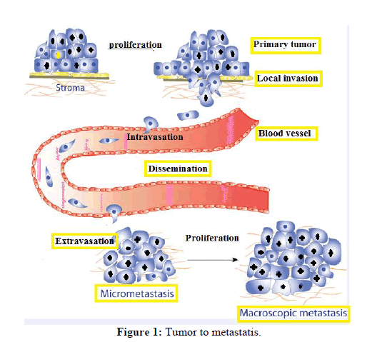
Cancer is a malignant disease that can be identified by uncontrollable proliferation of cells and unregulated cell expansion. It is still one of the world's most lethal weapons. According to the International Agency for Research on Cancer (IARC), 19.3 million new cancer cases will be determined in 2022, 10.0 million cancer-related deaths will occur and 28.4 million new cancer cases will be confirmed in 2030, a 47% increase from 2020. Lung, liver and stomach cancers were the leading causes of cancer deaths worldwide, followed by female breast cancer, lung and prostate cancer. Cancer passing away has gone down substantially in recent decades as early detection and treatment rates have increased. Furthermore, advances in molecular biology and tumour biology have been made. Over the last 15 years, cancer treatment methods have changed. Figure 1 depicts several cancer pathological features. Cancer cells' biological abilities include resistance to antigrowth signals, cell death resistance, unlimited replication possibility, inducing angiogenesis, active invasion and metastasis, deregulation of cellular energetics and immune destruction resistance. Tumor-associated inflammatory processes, which may deliver bioactive substances to the Tumour Microcosm (TME), as well as cancer cell genomic instability, are both required for the aforementioned features to be acquired. TME is a factor in growths that is integrated with neoplastic cells such as cancer stem cells, cancer-associated fibroblasts and cancer-associated fibroblasts. Endothelial cells, pericytes and immuneinflammatory cells are every kind of cells. Tumor-associated inflammatory processes, which can provide bioactive substances to the Tumour Microcosm (TME), as well as carcinoma cell genomic instability, are both required for getting hold of the aforementioned characteristics. TME is a tumour component that interacts with tumors stem cells, tumor-associated fibroblasts and cells of the blood vessels, pericytes and immuneinflammatory cells [1].
Bioactive compounds are becoming more credible as cancer preventative and therapeutic agents, according to growing evidence. The American Institute for Cancer Research (AICR) recently revised its list of 26 anticancer foods in its "global diet and cancer research update." In response to the report, strong evidence is defined as research that clearly establishes a causal link between cancer and the condition, while limited evidence indicates that, while the overall findings are usually backed by data, the evidence is rarely sufficient to support cancer risk reduction recommendations. Additionally, we will go into greater detail about the drugs' anticancer mechanisms in the following section. Finally, adjusting one's diet could prove able to halt many tumours' destructive processes. This review will start by going over some common bioactive substances before summarizing their effects on cancer and cancer-related pathways. The role of bioactive ingredients in cancer prevention is depicted in Table1.
| Bioactive compounds | Natural products | Against cancer |
|---|---|---|
| Triterpenoid compounds | Apples | Colorectal cancer |
| Vit c, chlorogenic acid, proanthocyanidins | Blue berries | Colorectal cancer |
| Flavonols, folate | Asparagus | Breast cancer |
| Carotenoids, lutein | Broccoli and cruciferous vegetables | Colorectal cancer |
| Carotenoids,phenolic acid | Carrots | Lung cancer |
| Anthocyanins, melatonin, phenolicacids | Cherries | Colon cancer |
| Terpenes, coumarins, flavanones | Grapefruit | Breast cancer |
| Flavonones | Orange | Stomach cancer |
| Ellagitannins,anthocyanins | Raspberries | Lung cancer |
| Stilbenes, resveratrol | Strawberries | Esophageal cancer |
| Lycopene, beta-carotene | Tomtatoes | Lung and stomach cancer |
Table 1: Bioactive compound against cancer.
Literature Review
Fruit derived bioactive compounds (Polyphenols)
Polyphenolic compounds are naturally occurring substances that have varying phenolic tasks. These substances are abundant in plants and play an important role in pathogen defense and cell growth control. Currently, numerous clinical studies are being conducted to determine how they are beneficial in the prevention of ageing, problems with metabolism and cardiovascular disorders. More research has focused on polyphenols' anticarcinogenic properties (such as reducing tumour expansion, metastasis and angiogenesis) as a result of a better understanding of these compounds. Polyphenolic chemicals are classified into four groups based on the number of phenol rings and molecular structures: phenolic acids, stilbenes, lignans and flavonoids. Table 2 shows examples and structures of phenolic acids [2].
| Phenolic compounds | Example | Structure |
|---|---|---|
| Flavanoids | Flavones, anthocyanine, isoflavanoids | 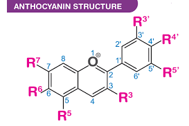 |
| Stilbenes | Resveratrol | 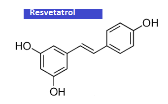 |
| Phenolic acids | Benzoic acid, cinnamic acid | 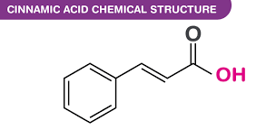 |
| Lignans | Matairesinol | 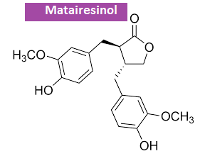 |
Table 2: List of phenolic acids with few example and structure.
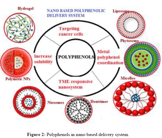
The majority of polyphenols present in foods exist as polymers, esters or glycosides; when polyphenols enter the body, bacteria in the intestines hydrolyze them before intake. Polyphenols then take part in methylation that exists sulfation and glucuronidation reactions. These polyphenolic compounds were found in bloodstream but not in organic foods. Absorption, metabolism and excretion rates are just a few of the factors that affect how polyphenols behave biologically. Furthermore, hepatic activity influences not only polyphenol metabolism but also the rate at which they enter cells and tissues. Polyphenols found in food have been shown to inhibit a variety of biochemical processes such as oxidation, tumour cell apoptosis, immune system activation and anti-inflammatory properties. More information on these and other biochemical processes will be provided in the future. Figure 2 express the nano based delivery of polyphenols [3].
Discussion
Effects of phenolic acid against various types of cancer
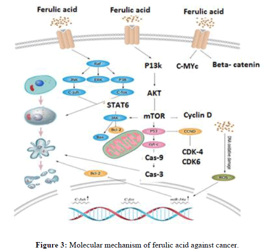
The Effects of bioactive compound ferulic acid, obtained from tomato on various types of cancers: The process by which FA and its byproducts kill cancer cells. FA promotes cell death by increasing P53 expression, decreasing cyclin D1 and CDK 4/6 expression, increasing Mir- 43a expression, suppressing Bcl-2 expression and activating the apoptotic pathway. FA causes DNA oxidative damage and apoptosis by producing ROS (figure 3). ERK, STAT signal transduction and transcriptional activator, C-fox forkhead box C, JAK the Janus kinase, Bcl-2-associated X (Bax), BCL-2 (B-cell lymphoma-2) and AKT protein kinase B are all names for JNK. JNK is also referred to as P53 (tumour protein 53) and caspase cysteinyl aspartate (Figure 3) [4,5].
Pharmacological role of quercetin
There are antioxidant qualities to quercetin. Antioxidants help cells resist stress caused by oxidative stress. When the body's defences against antioxidants are outnumbered by the body's excess free radicals, oxidative damage occurs. In healthcare, this is referred to as oxidative stress. Free radicals are erratic molecules in the body that can hasten ageing and increase the risk of disease. During routine metabolic functions such as energy synthesis, the body produces free radicals. Figure 5 depicts quercetin's therapeutic role [6].
Molecular mechanism of quercetin against cancer
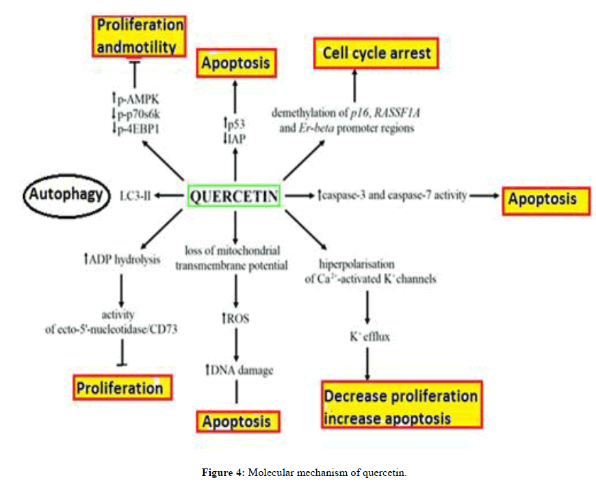
Quercetin increased DNA disintegration, subG0/G1 cell percentages and caspase 3/7 activity (Figure 4). Autophagy, on the other hand, could be to blame for the quercetin-induced decrease in cell viability. After quercetin treatment, the concentration of a specific autophagy marker, such as LC3- II, increased in human BC cells. Quercetin in combination with an autophagy inhibitor (bafilomycinA1), on the other hand, may be used to more effectively suppress the growth of bladder cancer cells by improving apoptosis. An in vivo study revealed that quercetin might lead BC cells to die by controlling p53. According to literature reviews, quercetin administration decreased the expression of the mutant P53 protein (mutP53). Furthermore, this treatment has been linked to a drop in surviving. It is also possible that quercetin's antiproliferative action on bladder transitional cell carcinoma is mediated, at least in part, by changes in nucleotide extracellular catabolism. These changes could be the result of AMP accumulation or quercetin-blocked adenosine receptors. (The very first) Ecto-50-nucleotidase catalyses AMP hydrolysis into adenosine, which has been linked to cancer progression, cell proliferation, maturation, differentiation, treatment resistance and tumour promotion. An in vivo study found that quercetin increased ADP hydrolysis, inhibited the activity of ecto-50-nucleotidase and CD73 and had no effect on protein production [7-9].
Effects of bioactive compounds obtained from pomegranate on various types of cancers
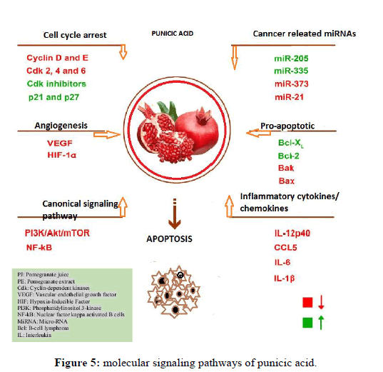
Punica granatum (Punica granatum): Pomegranate (Punica granatum L.), a member of the Lythraceae family, is a spherical berry with between 250 and 1500 white seeds that turn deep red or purple when ripe. Due to the presence of tannins and anthocyanins, seeds that are consumed have anti-inflammatory and antioxidant properties. Since ancient times, pomegranates have been used as medicine for a variety of ailments. It has antiparasitic properties and can help with stomach ulcers and diarrhoea. The Unani method of medicine, another conventional medical system, has even gained understanding for its effectiveness in the management of insulin resistance. Aside from the fruits, the bark, leaves, roots and other parts of the plant have been found to shown to be composed of huge quantities of molecular components with medicinal properties. It contains a bioactive substance that has been shown to influence a number of signalling pathways involved in inflammation, carcinogenesis, hyper proliferation, cellular transformation, angiogenesis and eventually, metastasis. The bioactive ingredients in pomegranate have been found to modulate a wide range of transcription factors, pro- and anti-apoptotic molecules, cell cycle-regulating molecules, protein kinases, cell adhesion molecules, proinflammatory mediators and growth factors [10].
Effect of punicic acid in various cancers
Punicic acid: Trichosanic acid or punicic acid, is a polyunsaturated fatty acid with an 18:3 cis-9, trans-11 and cis-13 ratio. It is made from pomegranate seed oil and bears the name of the pomegranate, Punica granatum. It has also been discovered in snake gourd seed oils.
Sun exposure is the leading cause of skin cancer. UVB light activates several signalling pathways in the skin, making it a risk factor for skin cancer (Sung H et al 2021 and Aqil F et al 2012). Pomegranate Fruit Extract (PFE), Pomegranate Juice (PJ) and Pomegranate Seed Oil (PSO) can all help prevent UVB-induced skin cancer. They've all been tested on skin cancer animal models, reconstituted human skin models and cell culture. In Normal Human Epidermal Keratinocytes (NHEK), PFE inhibited the phosphorylation of UVB-induced Mitogen-Activated Protein Kinases (MAPK) (Figure 5).
Breast cancer: According to reports, polyphenols from the PJ, pericarp and PSO inhibit the transformation of testosterone to oestrogen by preventing aromatase activity which may play an important part in breast cancer. In addition to anti-estrogenic attributes, these polyphenols were found to inhibit cell proliferation in the cell lines MCF-7 and MB-MDA-231. Another study discovered that pomegranate ellagitannin variations inhibit the formation of breast cancer cells. PFE and its components, according to studies, are extremely important for chemoprevention against breast cancer due to their pro-apoptotic and antioxidant properties. Punicic acid, which is found in PSO, has also been shown to induce apoptosis in the breast cancer cell lines MDAMB-231 and MDA-ER-7, both of which are estrogen-sensitive and insensitive [11].
Effect of caffeic acid against cancer
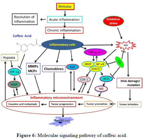
Caffeic acid: One type of chemical substance that falls under the hydroxycinnamic acid classification is caffeine. This yellow solid has functional groups that include acrylic and phenolic. Because it is an intermediary in the formation of lignin, one of the main constituents of the biomass and residues of woody plants, it is present in all plants (Figure 6).
CA has been shown to inhibit 5-lipoxygenase and decrease IL-6, IL-1 and NF-B in the course of inflammatory responses (Figure 6). CA suppresses STAT3 activity, which in turn reduces HIF-1 activity (Clinton et al, 2009). It contains potential STAT3 inhibiting agents and hampers tumour angiogenesis by inhibiting STAT3 activity as well as VEGF and HIF-1 expression. CA inhibited the phosphorylation of ERK as well as the transactivation of NF-B and AP-1. By targeting MEK1 and TOPK, CA, on the different hand, prevents tumour metastasis and neoplastic cell transformation. In fact, it connected with ERK1/2 in vitro and inhibited its activity [12].
Effect of ellagic acid against cancer
Ellagic acid: Ellagic Acid (EA) is a bioactive polyphenol produced naturally by many plant species as a secondary metabolite. Significant amounts of EA are found in the pomegranate (Punica granatum L.), as well as the wood and bark of several tree species. EA is a dilactone of the widely distributed class of secondary metabolites known as ellagitannins that is primarily formed by the hydrolysis of Hexahydroxydiphenic Acid (HHDP), a dimeric gallic acid derivative. EA's antioxidant, anti-inflammatory, antimutagenic and antiproliferative properties are attracting interest.
Molecular mechanism of ellagic acid against cancer
Ellagic acid, a benzopyranoid, is found in a variety of fruits and vegetables, including blackberries, cranberries, raspberries, strawberries, wolfberries, grapes, pomegranates, pecans and walnuts. According to studies, ellagic acid and its metabolites are anti-cancer. Signalling pathways are modulated specifically. Cell proliferation pathways (cyclin dependent kinase 2, cyclin A2, cyclin B1, cyclin D1, c-myc, PKCa), survival/apoptosis pathways (Bcl-XL, Bax, Caspase 9/3, Akt), angiogenesis pathways (VEGF) and inflammatory metastasis pathways are all involved (Figure 7) [13].
Effect of Emblica officinalis against cancer The Euphorbiaceae family's Emblica officinalis Gaertn or Emblic myrobalans, additionally referred to as Indian gooseberry in the United States and amla in Hindi, is a medium-sized deciduous tree. All parts of the plant have health advantages and are utilized for healing various medical conditions, but the fruit is extremely useful in many traditional systems of medicine and is used for a variety of culinary purposes. Amla is considered a powerful Rasayana or rejuvenator in Ayurveda because it helps to slow down the ageing process, promote longevity, benefit the eyes, improve digestion and treat constipation, prevent peptic ulcers, reduce fever and purify the blood, as well as having hepatoprotective, cardioprotective, diuretic and antiinflammatory properties. It is used to treat a wide range of conditions, including anaemia, hyperacidity, diarrhoea, eye inflammation, leucorrhea, jaundice, nerve debility, liver complaints and cough and hair loss. Amla contains a high concentration of vitamin C, tannins, alkaloids and phenolic compounds.
The fruit contains gallic acid, elagic acid and corilagin. Chebulinic acid, chebulagic acid, emblicanin a, emblicanin b, punigluconin, pedunculagin, citric acid, ellagitannin, trigalloyl glucose, pectin, 1-ogalloyl-b-d-glucose, 3,6-di-ogalloyl-d-glucose, corilagin, 1,6-di-o-galloyl-b-d-glucose, 3 ethylgallic acid (3 ethoxy 4,5 dihydroxy benzoic acid) and isostrictiniin along with flavonoids like quercetin, kaempfero etc.
The antiproliferative activity of amla extract was found to be very functional in human tumour cell lines of various histological origins, including human erythromyeloid K562, B-lymphoid Raji, T-lymphoid Jurkat, MCF7 and MDA-MB-231 breast cancer cell lines. In vitro, the aqueous extract triggered cytotoxicity in L 929 cells as well as apoptosis in Dalton's lymphoma ascites and CeHa cell lines. Normal and cancer cells differ primarily in their loss of distinctiveness, rather than indefinite replicative ability, increased discomfort and metastasis. In the in-vitro matrigel invasion assay, amla constituents prohibit metastasis and its liquid extract prevents MDAMB-231 cell invasion. The amla bioactive kaempferol has been shown to inhibit the expression of stromelysin 1 (MMP-3) in the MDA-MB-231 breast cancer cell line. Gallic acid, a polyphenol, has also been shown to inhibit the migration of gastric adenocarcinoma cells and the metastasis of P815 mastocytoma cells to the liver of DBA/2 mice.
Effect of gallic acid against cancer
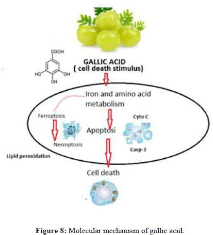
Gallic acid: Gallic acid, a naturally generated polyphenolic material, can be found in handled beverages such as red wine and green teas. It can be found in plants as hydrolysable tannins1-2, free acids, esters, catechin derivatives and free acids. The medicinal benefit of these compounds as radical scavengers has sparked the interest investigators. It has been shown to have potential therapeutic and preventive effects in a wide range of oxidative stress-related illnesses, which includes ageing, cardiovascular disease cancer and neurological disorders (Figure 8).
Gallic acid-induced cell death mechanisms have been proposed (Figure 8). Gallic acid activates the apoptotic, ferroptotic and necroptotic pathways, resulting in cell death. DFO, an iron chelator, has been shown to reduce certain types of cell death, proving that they are iron-dependent. Even when used in combination, inhibitors of three distinct cell death pathways, including the apoptosis inhibitor Z-VAD-FMK, ferroptosis inhibitors AOA and Fer-1 and necroptosis inhibitors Nec-1 and NSA, fail to prevent gallic acid-induced cell death. This discovery suggests that cell death may occur via an unidentified downstream pathway.
Effect of corilagin against cancer
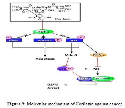
Corilagin: Corilagin, also known as 1-O-galloyl-3,6-(R)-hexahydroxydiphenoyl-d-glucose, is a naturally occurring Ellagitannin (ET) found in many plants. Corilagin has been shown to have antioxidant, anti-inflammatory, hepatoprotective, antibacterial, antihypertensive and anticancer properties. It has strong antistaphylococcal activity and inhibits RNA tumour virus reverse transcriptase activity (Figure 9). ET's antiproliferative effect has been attributed to a number of mechanisms, including cell cycle inhibition, mitochondrial apoptosis and self-destruction after replication.
TGF-isoforms cause cancer cells to secrete pro-Matrix Metalloproteinase (MMP), which results in cell-to-cell contact loss, increased N-cadherin communication, reduced E-cadherin expression and the development of a fibroblastoid phenotype. All of these processes match to the epithelialmesenchymal transition. TGF- is also linked to receptors of type I (ThRI) and type II (ThRII). After binding to its ligands, ThRII is activated, resulting in the phosphorylation in of receptor-regulated Smads. Smads that have been phosphorylated bind to co-Smads and Smad4. The movement of this complex changes gene expression in the nucleus. TGF- activates PI3K and MAPK family members such as c-Jun-NH2-kinase, TAK1, p38 and extracellular signal-regulated kinase. Corilagin may stop the cell cycle in the S- and G2/M phases by inhibiting the cyclin B1/cdc2 complex which is crucial for controlling the transition from the G2 to the M phase in SKOv3ip cells [14].
Effect of p-coumaric acid against cancer
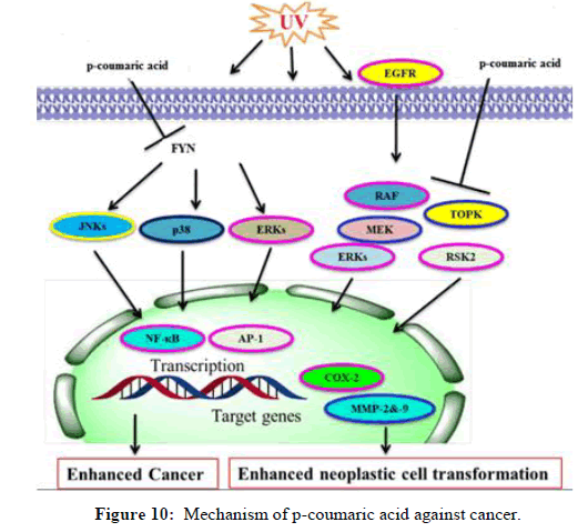
p-coumaric acid: When it comes to inhibiting Fyn kinase and suppressing the activation of MAPK pathways, CA is more effective than chlorogenic acid. Nonetheless, B-Raf mutations are frequently seen in colon polyps and are a precursor to both colon cancer and melanoma. MEK1/2 is distinct from the other elements of MAPK pathways. In rodent models and cell culture, constitutive MEK1 activation influences transformation and cancer, while an inhibitor of MEKs suppresses these processes. The kinase known as lymphokine-activated killer TOPK is known to activate ERKs and TOPK activation is implicated in the carcinogenic role of TOPK in malignancies. Through inhibiting ERKs phosphorylation, CA inhibited neoplastic cell transformation and CT-26 cell-mediated lung metastasis in mice. CA or decaffeinated coffee inhibited the phosphorylation of ERKs and the actions of COX-2, MMP-2 and -9 mediated by CT-26 cells (251, 252). Results from laboratory trials and computational modeling studies demonstrated that CA specifically interacted with mitogen-activated MEK1 and TOPK with ATP, repressing their respective kinase activities. Attenuation of AP-1 and NF-κB transactivation, TPA-mediated ERKs and p90 RSK2 and suppression of TOPK and MEK1 have all been related. Reduced ERK phosphorylation in colon cancer has been associated with CA/coffee consumption. Nonetheless, a summary of the CA effects and molecular targets is provided. According to ERK/Nrf2 signaling, CA was shown to be a stimulant of HO-1, GCLC and GCLM. As such, it may be a useful chemoprotective medication to protect liver injury from oxidative damage. Chemo preventive action is taken by CA against cancer (Figure 10).
Conclusion
Cancer is the most common cause of illness and death on a global scale. It is critical to develop more effective and targeted treatment plans to halt the progression of cancer. Biologically active substances have been identified as cancer treatments the fact double as chemotherapeutic and chemopreventive. Furthermore, it has been proposed that the effect of bioactive chemicals on cancer may be impacted by a number of mechanisms, including ROS, ER stress and epigenetic changes. More experiments will be conducted with the goal to better understand the mechanisms that underlie the anticancer effects. This information may be useful in developing synergistic combinations to improve efficacy.
References
- Bray F, Ren JS, Masuyer E, et al. Int J Cancer Res. 2013; 132(5): p. 1133-1145.
[Crossref] [Google Scholar] [PubMed]
- Roy A, Datta S, Bhatia KS, et al. S Afr J Bot. 2022; 149: p. 1017-1028.
- Singh N, Yadav SS. Curr Res Food Sci. 2022; 5: p. 1508-1523.
[Crossref] [Google Scholar] [PubMed]
- Gregoriou G, Neophytou CM, Vasincu A, et al. Molecules. 2021; 26(16): p. 5017.
[Crossref] [Google Scholar] [PubMed]
- Baloghová J, Michalkova R, Baranova Z, et al. Molecules. 2023; 28(17): p. 6251.
[Crossref] [Google Scholar] [PubMed]
- Samec M, Liskova A, Kubatka P, et al. J Cancer Res Clin Oncol. 2019; 145: p. 1665-1679.
[Crossref] [Google Scholar] [PubMed]
- Eskra JN, Dodge A, Schlicht MJ, et al. Nutr Cancer. 2020; 72(4): p. 672-685.
[Crossref] [Google Scholar] [PubMed]
- Asma ST, Acaroz U, Imre K, et al. Cancers. 2022; 14(24): p. 6203.
[Crossref] [Google Scholar] [PubMed]
- Garbe C, Amaral T, Peris K, et al. Eur J Cancer. 2022; 170: p. 256-284.
[Crossref] [Google Scholar] [PubMed]
- de S, Paul S, Manna A, et al. Cancers. 2023; 15(3): p. 993.
[Crossref] [Google Scholar] [PubMed]
- Menter T, Tzankov A, Dirnhofer S. Hem Oncol. 2020; 39(1): p. 3-10.
[Crossref] [Google Scholar] [PubMed]
- Moeini S, Karimi E, Oskoueian E. Cancer Nanotechnol. 2022; 13(1): p. 20.
- Subramaniam S, Selvaduray KR, Radhakrishnan AK. Biomolecules. 2019; 9(12): p. 758.
[Crossref] [Google Scholar] [PubMed]
- Logan J, Bourassa MW. Eur J Cancer Preven. 2018; 27(4): p. 406-410.
[Crossref] [Google Scholar] [PubMed]

