Research Article - Der Pharma Chemica ( 2021) Volume 13, Issue 8
Synthesis, Spectral Elucidation, Antibacterial, Antioxidant and DNA Studies Of ONNO Tetradentate Schiff Base Metal(II) Complexes derived From Benzene-1,4-Dicarboxaldehyde
Kumarasamy Savitha1 and SubramaniamVedanayaki1*SubramaniamVedanayaki, Department of Chemistry, Kandaswami Kandar’s College, ParamathiVelur(Tk), Namakkal(Dt), TamilNadu- 638182, India, Email: varshuvishal@gmail.com
Received: 19-Jan-2021 Accepted Date: Aug 07, 2021 ; Published: 14-Aug-2021
Abstract
The new dibasic tetra dentate (ONNO) Schiff base ligands (L1and L2) were synthesized by the reaction of terephthalaldehyde with 2-amino 4-chlorophenol and X (where, X=2-amino 4-methylphenol, 2-aminophenol) in 1:1:1 molar ratio. The macrocyclic binuclear Co (II), Ni (II) and Cu ((II) metal complexes were prepared in ligand to metal ratio 2:2. The elements, metal structure and binding sites of Schiff bases and its complexes were established by diverse studies like Elemental, Molar conductance, UV, Magnetic moment, FT-IR, H1 and C13 NMR, ESI-Mass, ESR, Thermal and Powder-XRD. The above spectral studies reveal that the ligands are a tetra dentate and its metal complexes possess a square planar geometry. All the compounds were screened for antibacterial, antioxidant and DNA cleavage and the results show high activity for metal complexes than the ligand. The DNA binding studies of Cu(II) complexes were measured by electronic absorption method. In vitro cytotoxicity assay of Cu (II) complexes were tested for their tumour inhibiting potential against MCF-7 human breast cancer cell by using MTT method.
Keywords
Schiff bases; Square planar; Antimicrobial; Antioxidant; DNA studies; Antitumour activity
Introduction
Chelating Schiff base having mono, di and polydentate donor atoms of O, N and S shows biological activity. It is bound to metal ions and Schiff base metal complexes give the small changes in the geometry which depends upon its ligand having hard and soft donor atoms [1,2]. N2O2, NNNN and ONO donor atoms of polydentate ligands give stable complexes with transition metal ions due to their close proximity of the donor sites and it gives four, five and six member chelate rings. Symmetrical diimines (~N=HC-Ar-CH=N~ (or) ~HC=N-Ar-N=CH~) formed from the condensation of one mole of an aromatic diamine and two moles of aldehyde (or) one mole of an aromatic dialdehyde and two moles of primary amine was prepared directly. Schiff base including two different imino groups attached to the same aromatic ring form an asymmetric diimine (~N=HC-Ar-N=CH~). Recently many literatures concerning synthesis of asymmetrical diimines have been published [3]. Biological applications of Schiff base complexes are one of the most completely studied topics in coordination chemistry due to its best activities than the non-schiff base metal complexes [4, 5]. Schiff base metal complexes have an extensively diverse structure from the coordination of metal with mono-, bi-, tri and tetra dentate ligand which is depends upon the carbonyl groups and amines. The symmetrical tetra dentate ligand complexes have been used as catalyst in some chemical process and suggested as very useful biological kinds in understanding no regular interaction of peptides [6]. Schiff base and their complexes indicate various possible mode of interaction, which is very importantly, both of aldimine- and ketimine- types of large. Schiff base ligands were given to shift small changes in its structure can affect significantly their antiproliferactive activity and considerable cytotoxicity activities due to its varied structure [7]. Schiff base transition metal complexes has more strong properties of structural, chemical and spectral character depends upon the nature of its ligand, which are particular attention to bio-inorganic and inorganic chemistry [8, 9].
In recent years, metal-based drugs act very important role in medicinal field, which are used as drugs for the cancer treatment, diabetes, cardiovascular diseases and anti-inflammatory [10, 11]. Transition metal complexes of 2-aminophenol based compounds have vast application fields in antidepressants, antiphlogogistic, nematocide and other medicinal agents [12]. Schiff base complexes are widely studied due to synthetic flexibility, selective and sensitivity towards a diversity of organisms, which these complexes have more potential application in more fields like as oxidation catalysis, analytical and electrochemistry, dye & food industry, agrochemical and pharmaceutical fields [13-15]. Intramolecular hydrogen bonding between hydroxyl group (OH) hydrogen and imino group (CH=N) nitrogen atoms of the Schiff base and their metal complexes plays a considerable role in many fields of anti-tubercular, anti-HIV, anthelmintic, anti-amoebic, antinociceptive, antimouse hepatitis virus (MHv), inhibition of herpes simplex virus type-1(HSV-1), adenovirus type-5 (AD-5), antimalarial, pesticidal, herbicidal, antipyretic, antiviral, antioxidant, antibacterial and antifungal [16-23]. Moreover, copper metal is a very important between other transition metals and it is the third most abundant metallic element in human body following the metals of iron and zinc. It plays many roles in the enzyme catalysis, necessary for the growth, maintenance and development of bones, connectivity tissues, heart, brain and many other body organs [24].
Based on the above fact, herein we have reported the synthesis of new asymmetric Schiff base ligands (4-chloro-2- ((E)- (4- ((E)- ((2-hydroxy-5-methylphenyl) imino) methyl) benzylidene) amino) phenol) (L1) and 4-chloro-2-((E)- (4- ((E)- ((2 hydroxyphenyl) imino) methyl) benzylidene) aminophenol (L2) containing the azomethine (CH=N) and hydroxyl groups (OH) as potent chelating sites and its Co(II), Ni(II) and Cu(II) binuclear metal complexes evaluated by diverse physicochemical technique and their applications were analyzed for antibacterial, antioxidant, DNA studies (cleavage and binding) and antitumour activity (in vitro cytotoxicity).
Experimental Section
Analytical and Physical Measurements
Terephthalaldehyde, 2-amino 4-chlorophenol, 2-amino 4-methylphenol, 2-aminophenol and metal salts were purchased from Sigma Aldrich. Ethanol, Methanol, DMSO, DMF and Acetone were purchased from Loba and Merck chemicals. The purity of all compounds was tested by TLC.
C, H and N elements was determined on a Thermo Finningan Flash EA 1112 series elemental analyzer. Molar conductance of the compounds was measured by ELICO CM 180 Conductivity Bridge. Magnectic moment values were observed using Gauy balance calibrated with Hg[Co(SCN)4] experiment. Electronic absorption spectrum was measured using Perkin Elmer Lambda -25 spectrometer. FT-IR spectra were recorded on a Shimadzu FT-IR-8300 spectrometer using KBr Pellet technique. H1 and C13-NMR spectra were obtained on a BRUKER ADVANCED III 400MHz spectrometer using TMS used as reference. ESI-Mass spectra were recorded on a Perkin- Elmer R MU-6E instrument. ESR spectra were measured on JES-FA200 ESR spectrometer with X-band freqenucy. Thermal analysis studies were carried out at 0˚-1000˚C using SDT-Q600 V20.9 Build 20 thermal analyzer. The P-XRD was recorded on Perkin Elmer TA/SDT-2960 and Philips 3701 instrument.
Synthesis of asymmetrical tetra dentate Schiff base L1 (compound 1)
The mixture of 1mmole of terephthalaldehyde with 1mmole of 2-amino 4-chlorophenol and 1mmole of 2-amino 4-methylphenol were dissolved in methanol. The solution was kept under stirring for 2 hrs, the formed yellow precipitate was separated by filtration, washed and purified by methanol solvent. The Schiff base solid was recrystalized from ethanol. Yellow solid, Molecular weight- 364.82, Melting point-240 ˚C, Yield- 80%, IR (KBr cm-1): 3345 (OH), 1621 (C=N), 1278 (C-O); 1H-NMR (DMSO-d6, δ, ppm): 8.75 (s, 1H, C=N), 10.1 (s, 1H, OH), 3.4(s, 3H, CH3), 6.9-8.2 (m, Ar—H); 13C-NMR (100MHz, CDCl3): 112, 113, 114, 116, 120, 124, 129, 135, 137, 147, 161; Elemental analysis: C21H17ClN2O2 Calculated values: C- 69.14, H- 4.70, N- 7.68: Found values: C- 68.86, H- 4.50, N- 7.98; ESI-Mass: m/z: (M+1)+ 365.
Synthesis of asymmetrical tetra dentate Schiff base L2 (compound 5)
The procedure is the same as L1 using terephthalaldehyde with 2-amino 4-chlorophenol and 2-aminophenol. The dark yellow precipitate was formed. Molecular weight- 350.08, Melting point-250 ˚C, Yield- 80%, IR (KBr cm-1): 3346 (OH), 1621 (C=N), 1369 (C-O); 1H-NMR (DMSO-d6, δ, ppm): 8.80 (s, 1H, C=N), 9.4 (s, 1H, OH), 6.8-7.3 (m, Ar—H); 13C-NMR (100MHz, CDCl3): 110, 115, 116,120, 123, 124, 126, 127, 128,130, 137, 138, 148, 150, 152; Elemental analysis: C20H15ClN2O2 Calculated values: C- 68.48, H- 4.31, N- 7.99: Found values: C- 67.71, H- 4.13, N- 8.12; ESI-Mass: m/z: (M+1)+ 351.
Synthesis of Binuclear metal (II) complexes
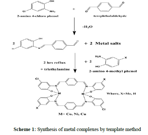
The homo binuclear metal (II) complexes were synthesized by using template method [25]. The mixture of terephthalaldehyde (2mmole) with 2-amino 4-chlorophenol (2mmole) and X (where, X=2-amino 4-methylphenol, 2-aminophenol) (2mmole) were dissolved in methanol, which were added to the methanolic solution of metal salts (2mmole) like Co, Ni and Cu. The mixture was stirred and few drops of tri ethylamine were added to the mixture. It was stirred for 1 hr and under reflux for 3 hrs. The product was partly evaporated, cooled at room temperature. The obtained metal complexes were separated by filtration, washed with methanol and diethyl ether, stored in room temperature. The structure of metal complexes was show in scheme 1.
Schiff base binuclear Co (II) complex (compound 2)
Brownish black solid, Molecular weight- 903.62, Melting point->300 ˚C, Yield- 78%, IR (KBr cm-1): 1606 (C=N), 1384 (C-O), 518 (M-O), 492 (M-N); Elemental analysis: C46H42Cl2Co2N4O4 Calculated values: C- 61.14, H- 4.68, N- 6.20, M- 13.04: Found values: C- 60.80, H- 4.51, N-6.18, M-12.98; Molar conductance ( Ω-1 cm2 mol-1) 10.6.
Schiff base binuclear Ni (II) complex (compound 3)
Reddish yellow solid, Molecular weight- 903.14, Melting point->300 ˚C, Yield- 76%, IR (KBr cm-1): 1565 (C=N), 1385 (C-O), 515 (M-O), 467 (M-N); Elemental analysis: C46H42Cl2N4Ni2O4 Calculated values: C- 61.17, H- 4.69, N- 6.20, M- 13.00: Found values: C- 61.12, H- 4.53, N- 6.21, M-12.81; Molar conductance ( -1 cm2 mol-1) 11.3.
Schiff base binuclear Cu (II) complex (compound 4)
Black solid, Molecular weight- 912.85, Melting point->300 ˚C, Yield- 79%, IR (KBr cm-1): 1601 (C=N), 1397 (C-O), 513 (M-O), 462 (M-N); Elemental analysis: C46H42Cl2Cu2N4O4 Calculated values: C- 60.52, H- 4.64, N- 6.14, M- 13.92: Found values: C- 60.35, H- 4.55, N- 6.04 , M- 13.82; Molar conductance ( -1 cm2 mol-1) 13.5.
Schiff base binuclear Co (II) complex (compound 6)
Wine red solid, Molecular weight- 875.57, Melting point->300 ˚C, Yield- 79%, IR (KBr cm-1): 1601 (C=N), 1385 (C-O), 583 (M-O), 468 (M-N); Elemental analysis: C44H38Cl2Co2N4O4 Calculated values: C- 60.36, H- 4.37, N- 6.40, M- 13.46: Found values: C- 59.81, H- 4.17, N-6.30, M-13.25; Molar conductance ( -1 cm2 mol-1) 10.3.
Schiff base binuclear Ni (II) complex (compound 7)
Yellowish brown solid, Molecular weight- 875.09, Melting point->300 ˚C, Yield- 75%, IR (KBr cm-1): 1583 (C=N), 1383 (C-O), 598 (M-O), 443 (M-N); Elemental analysis: C44H38Cl2N4Ni2O4 Calculated values: C- 60.39, H- 4.38, N- 6.40, M- 13.41: Found values: C- 60.15, H- 4.19, N- 6.30, M-13.19; Molar conductance ( -1 cm2 mol-1) 11.7.
Schiff base binuclear Cu (II) complex (compound 8)
Black solid, Molecular weight- 884.79, Melting point->300 ˚C, Yield- 78%, IR (KBr cm-1): 1572 (C=N), 1394 (C-O), 581 (M-O), 477 (M-N); Elemental analysis: C44H38Cl2Cu2N4O4 Calculated values: C- 58.73, H- 4.33, N- 6.33, M- 14.36: Found values: C- 58.82, H- 4.22, N- 6.21 , M- 13.10; Molar conductance ( Ω-1 cm2 mol-1) 13.8.
Pharmacological Studies
Antibacterial activity
In vitro antibacterial activity was performed by the Disc-agar well diffusion experiment. The asymmetric tetra dentate Schiff base ligands and their homo binuclear complexes were screened against bacteria of Gram-positive (S.aureus, B.subtillis) and Gram-negative (K.neumoniae, E.coli). Tetracycline was used as standard drug for antibacterial activity and the tested compounds were dissolved in DMSO. The antibacterial activities were maintained in nutrient agar well plates at 4˚C. The 100 μL concentration of culture supernant was placed on agar well plate and then it was incubated for 24 hrs at 37˚C. The antibacterial activity was determined by measuring the diameter of the zone indicating complete inhibition [26].
Antioxidant activity
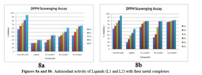
Schiff base Ligands (compound 1, 5) and their metal (compds.2, 3, 4, 6, 7, 8) complexes were investigated by using its free radical scavenging activity on the stable DPPH free radicals described in the literature [27]. The scavenging activity investigates the antiradical power of an antioxidant by calculating the decrease in the absorbance wavelength of DPPH at 510 nm. The ligands and its metal complexes exhibit DPPH free radical scavenging activity at different concentrations (like as 10, 20, 30, 40 and 50 μL). The percentage of DPPH free radical scavenging ability was measured by using the following formula
I% = Ac – As/ Ac X100
Where Ac is the Absorbance of the control and as is the Absorbance of the tested sample. IC50 values were calculated for ligand and its complexes, which showed the significant ability and it is defined as concentration sufficient to generate 50% of maximum scavenging activity. Ascorbic acid was used as standard.
DNA Binding Studies
Electronic Absorption Spectroscopy
Electron absorption spectroscopy is one of the most useful techniques to determine the interaction between the metal complexes with DNA from the stock solution of calf thymus (CT) [28]. DNA was prepared by 5mM Tris-HCL/20mM NaCl buffer (pH=7.2) at room temperature. The stock solution was stored at 4˚C and used up to 4 days. The buffer solution of CT- DNA gave a ratio of ~1.8-1.9 and the UV absorbance at 260 to 280 nm. The concentration of CT-DNA was determined using the known molar extinction coefficient value of 6600 M-1 cm-1 at absorption intensity is 260 nm. The stock solution concentrations of the complexes were prepared by dissolving the Cu (II) complexes (compds 4,8) in DMSO solvent. All the experiments were maintained at proper dilution with corresponding buffer to the required concentration. The binding constant (Kb) was calculated by using the equation:
[DNA]/ (εa-εf) = [DNA]/ (εa-εf) +1/Kb (εa-εf)
Where, [DNA] is molar concentration of DNA is base pairs, εa, εf are apparent extinction coefficient, the Kb values were obtained from the ratio between the equation of DNA/ (εa-εf) versus [DNA] in each case.
Agarose Gel Electrophoresis Assay
The cleavage of super coiled pBR322 DNA was investigated by gel electrophoresis method [29]. The agarose gel electrophoresis studies were carried out incubation of the mixtures containing 20 μL pBR322 DNA, 50 mM of NaCl, 50 mM of metal complexes and 50 mM H2O2 in Tris-HCl buffer (pH=7.4) at 37˚C for 1 hr. After incubation, the sample compounds were electrophoresed at 60˚C for 2 hrs on 1% of agarose gel using TAE (Tris-Acetic acid-EDTA) buffer (pH=8.0). After 0.5 μg/ml of ethidium bromide was used, the gel was stained. Thus all the experiments were performed at room temperature and photographed under UV light at 360 nm.
Antitumor activity
In vitro cytotoxicity was carried out on a MCF-7 cell line in which cell viability was tested using MTT (3-(4, 5-dimethyl thiazol-2-yl)-2,5-diphenyltetrazolium bromide) activity [30]. In vitro effect of growth inhibition of Cu (II) complexes (4, 8) were measured by spectrophotometric experiment. This experiment was used to determine the MTT conversion into “formazan” by living cells and it was maintained in a 96-well microplate having DMEM/F12 plain media supplemented with 10% heat inactivated FBS (Fetal Bovine Serum) having 5%of mixture of 100 μg/mL of streptomycin and 100 units/ml of penicillin were incubated at 37˚C for 24 hrs in the presence of 5% CO2. In this study, various concentrations of 10, 20, 40, 80, 160 and 320 μL of the stock solution was prepared in DMSO, which were added to relavant wells having 100 μL of the DMSO medium. After incubation, 100 μL of MTT stock solution (5mg/10ml of MTT in PBS) was added to each well and further incubated for 4 hrs. After the supernatant was removed, the plates were gently shaken to form solubilize formazan crystals by adding 100 μL of DMSO. The absorbance was measured at wavelength 590 nm by microplate reader. The activity was performed in triplicate and used to calculate the mean.
Results and Discussion
The synthesized ligands and their metal complexes were investigated by diverse physicochemical methods. The resultant compounds are soluble in DMF and DMSO, partially soluble in ethanol and methanol.
Elemental and Molar conductivity analysis
The composition of C, H and N are analyzed by CHNS elemental analyzer. The molar conductance of ligands and their complexes were measured at 25˚C in DMF solution (10-3M) indicates low conductance values (10.3 to 13.8), which shows that the complexes are non-electrolytic nature [31, 32].
Electronic absorption spectral data
The UV-Vis spectral data of the ligand and its complexes were measured within a 200-800 nm wavelength at room temperature in DMSO solution. In the electronic spectra of the mixed ligands (L1 and L2) were exhibit absorption bands at 300, 299 and 392, 388 nm, which is referred to the aromatic benzene rings (π→π*) and imine group (nrespectiv ely [33].
The diffuse reflectance spectrum of the macrocyclic metal(II) compounds of 2, 3,4, 6, 7 and 8 show bands at 442, 439, 440, 436, 414 and 438 nm, which may be attributed to the ligand to metal co-ordinations (L→M transitions) in square planar geometry of the compounds (Table 1).
| S.No | Compounds | n | LM | Geometry | µeff (BM) | |
|---|---|---|---|---|---|---|
| 1 | [L1] | 300 | 392 | --- | --- | --- |
| 2 | [Co2L2] | 251 | 374 | 442 | Square planar | 1.82 |
| 3 | [Ni2L2] | 253 | 370 | 439 | Square planar | --- |
| 4 | [Cu2L2] | 252 | 378 | 440 | Square planar | 1.71 |
| 5 | [L2] | 299 | 388 | --- | --- | --- |
| 6 | [Co2L2] | 271 | 354 | 436 | Square planar | 1.80 |
| 7 | [Ni2L2] | 251 | 370 | 414 | Square planar | --- |
| 8 | [Cu2L2] | 279 | 369 | 438 | Square planar | 1.76 |
The Traditional medicine also known as alternative, complimentary, and indigenous or folk medicine comprises knowledge systems that developed over generations within various societies before the era of modern medicine.
FT-IR studies
IR spectrum is used to investigate of the existence of intra molecular hydrogen bonding, the nature of the co-ordination mode in the metal complexes and presence of the tautomeric forms in the solid states. The FT-IR spectral data of the ligands and their complexes were observed in the wave number region at 400-4000 cm-1. The important vibrational frequency of the free ligands (L1 and L2) were exhibits band at 1621 and 1621 cm-1, which indicates the formation of imine groups(C=N) [35]. However, on complexation this band(C=N) is shifted to lower energy range 1606-1565 cm-1, suggesting the participation of the azomethine nitrogen atom in co-ordination to the metal ion. The sharp broad bands around 3345 and 3346 cm-1 in the ligands (L1 and L2) were assigned to the phenolic –OH groups. The absence of ν (OH) bands in the metal complexes, indicates the co-ordination of the ν(OH) groups after deprotonation. The ν(C=O) band at 1278 and 1369 cm-1 for L1 and L2 were lower frequency than the band at about 1700 cm-1 for ν(C=O) , this frequency change shows the formation of azomethine group(ligand).
The appearance of new bands at 443-492 and 513-598 cm-1 in the vibrational spectra of the complexes were assigned to ν(M-N) and ν(M-O) frequencies respectively [36]. These vibrations were clearly suggesting the co-ordination of the metal ions with the azomethine nitrogen and phenolic oxygens in the complex.
NMR Spectral Studies
1H-NMR spectrum
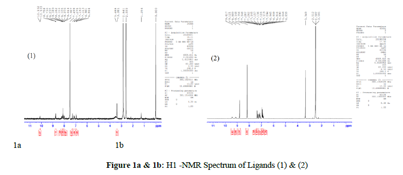
The 1H NMR spectrum of the ligands of L1 and L2 were recorded at 25˚C (RT) in DMSO-d6 solvent with TMS as internal standard. The azomethine (CH=N) proton signal was obtained at δ8.75 and δ8.80 ppm, this signal indicates the formation of mixed Schiff base ligands (L1 and L2) (shown in Figures 1a and 1b for Ligands (1) and(2)) [37]. The new peaks of phenolic –OH protons of the aminophenol and aromatic protons were observed at δ10.1, δ 9.4 ppm, and δ6.9-8.2, δ 6.8-7.3 ppm [38].
13C-NMR spectrum
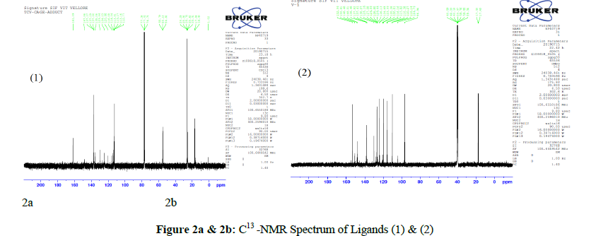
13C- NMR spectrums of the synthesized ligands (L1 and L2) were shifted to δ 161and δ152 ppm for azomethine carbon atom. The peaks showed at δ 147, δ 150 ppm and δ 112-129, δ 137-110 ppm were due to phenolic –OH carbons by aminophenol and aromatic benzene ring carbon atoms for schiff base Ligands (1) and (2) are shown in Figures 2a and 2b. The peaks appeared at δ137, δ138 ppm were due to substituted Cl atoms by aminophenol for L1 and L2. The signal appeared at δ 135 ppm was referred to the methyl group of the 2-aminophenol moiety for L1 [39].
ESI-Mass spectrum
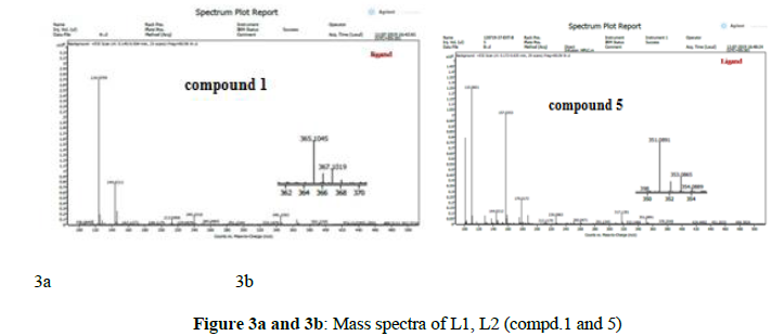
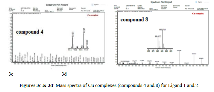
The electronic impact spectrum of Schiff base ligands (L1, L2) and their copper complexes (compouds 4, 8) showed molecular ion peaks at m/z 365, m/z 351 (M+1)+ and m/z 913, m/z 885 (M+1)+ , which corresponds to the proposed molecular formula of ligands L1 (C21H17ClN2O2), L2 (C20H15ClN2O2) and their Cu complexes 4 (C46H42Cl2Cu2N4O4), 8 (C44H38Cl2Cu2N4O4) respectively [40]. The mass spectral data of Schiff base ligands(L1 and L2) and their copper (II) complexes(compounds 4, 8) were confirmed by comparing their molecular formula weights with (m/z) mass values, which is in good agreement for these ligands ( compounds 1 and 5) and Cu( compounds 4 and 8) complexes as shown in figures 3a, 3b, 3c, 3d.
Electron Resonance Spectrum
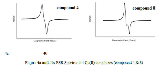
The electron spin resonance studies of the binuclear Cu (II) complexes of compounds 4, 8 gives information about hyperfine and superhyperfine structures and about the nature of the bonding between the copper ion and their ligand [41]. The EPR spectrum of complexes compd. 4 (Cu2L2) and compd. 8(Cu2L2) were displayed at room temperature and which exhibits an axial symmetry at X-band frequencies in the solid state. The obtained g-values are gII=2.2391, g⟘=2.0554 and gII=2.2418, g⟘=2.0498 for compounds 4 and 8, which were related by G=(gII-2)/(g⟘-2)=4.0. If G value is more than 4 ie(G>4), which indicates no considerable exchanging interaction between the two copper centers in the complex. Whereas if G value is less than 4(G<4), which shows that the exchange interaction occurs in the solid state complex. From the observed G value of Cu(II) complexes are 4.31 and 4.85 for compounds 4 and 8. It is clear that gII>g⟘>2.0023, which indicates dx2-y2 in orbital ground state for square planar structure of the Cu(II) complexes 4 and 8 shown in figures 4a and 4b.
Thermal studies
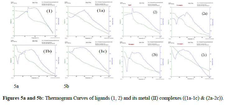
The thermal analysis (TG & DSC) was used to determine thermal stability of compounds for ligands and their metal complexes in the presence of air atmosphere at the temperature range between 0 to 1000°C [42]. Thermogram of the ligands (L1 and L2) and their complexes (2, 3, 4, 6, 7and 8) were shown in Figures 5a and 5b. The both TG and DSC analysis of compounds have three steps of decomposition process each. The first, second and third steps correspond to the elimination of small groups of substituted Cl atoms by 2-aminophenol, the removal of total ligand moiety and the formation of metal oxide residue.
P-XRD analysis
The X-ray powder diffraction analysis of the synthesized compounds of ligands and their metal complexes has been carried out to confirm whether the nature of the sample is amorphous (or) crystalline. The structure of Schiff bases and their binuclear metal complexes were investigated using P-XRD analysis which indicates the amorphous orthorhombic crystal nature of compounds within the range 10-90˚C (2θ) and the wavelength is 1.5406 A˚ [43]. Thus all the compounds show crystalline nature from its observed peaks. The Powder-XRD provides the d-value, relative intensity and 2θ value for each peak. The X-ray powder diffraction of ligands and their metal complexes are shown in figures 6a, 6b.
Biological Applications
Antibacterial assay
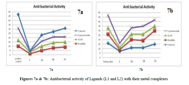
The antibacterial activity of tetra dentate Schiff base ligands (compounds 1 and 2) and their metal complexes (compounds 2, 3, 4, 6, 7, 8) were screened against for S.aureus, B.subtillis (gram-positive) and K.pneumoniae, E.coli (gram-negative) bacteria and tetracycline was used as standard drug. The antibacterial results showed that the Schiff base ligands have very low (or) no inhibition activity compared to their metal complexes [44]. Thus Cu(II) complexes (compounds 4, 8) were exhibits higher antibacterial activity than the other Co(II) (compounds 2, 6) and Ni(II) (compounds 3, 7) complexes. Antibacterial activities of L1 and L2 with their metal complexes are shown in figure 7a and 7b.
Antioxidant assay
DPPH free radical scavenging activity
The ligands (compounds 1, 2) and their Co(II) (compounds 2,6), Ni(II) (compounds 3, 7) and Cu(II) (compounds 4, 8) complexes were screened for their DPPH free radical scavenging ability using Ascorbic acid as standard [45]. The ligands have very low activity when compared to all the complexes. The scavenging activity of Cu (II) complexes are higher than that of other metal complexes. The IC50 values were determined (shown in figures 8a, 8b) for all compounds and the IC50 values of [Cu2 (C46H42N4O4Cl2)] and [Cu2 (C44H38N4O4Cl2)] were showed significant activity compared to other complexes and ligands. The order of scavenging activity of all complexes according to their IC50 values is given below.
Ascorbic acid > [Cu (II) complexes] > [Ni(II) complexes]> [Co(II) complexes]> Ligands Figures
DNA- copper complex interaction studies
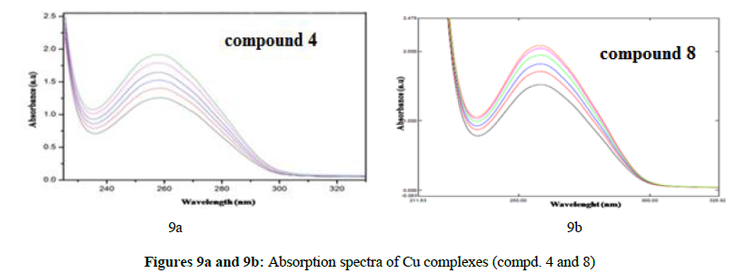
The interactions of CT-DNA and metal complexes (4, 8) were determined by the electronic absorption spectroscopy technique. From the studies it is revealed that the intensity changes of the intraligand π → π* transition band occur at 250-280 nm [46]. The UV absorption experiments of Cu (II) complexes (compounds 4, 8) in presence of buffer solution are performed by using a fixed concentration of complex to which increments of the stock solution was added. The interaction of [Cu2 (C46H42N4O4Cl2)] and [Cu2 (C44H38N4O4Cl2)] complexes with duplex DNA led to a decrease in the intensities and a small amount of red shift in the electronic absorption spectra and the concentration of DNA increases with the absorption bands of Cu(II) complexes were affected to a considerable extent. This absorption spectra indicate clearly that the addition of CT-DNA to the Cu (II) complexes yields hypochromic and red shift (shown in compound 8) to the ratio of [DNA]/[Cu] for the compounds 4, 8 complexes and the complexes interact with CT-DNA most likely through a binding mode that involves π → π* stacking interaction between the aromatic chromophore and the base pair of DNA
H2O2 induced DNA damage/ production assay
Gel electrophoresis activity is a method to determine different binding modes of newly synthesized complexes to super coiled pBR322 DNA. The natural- derived plasmid pBR322 DNA has three forms of closed-circle super coiled form (form-I), nicked form (form-II) and linear form (form-III) [47]. The circular plasmid DNA is conducted by electrophoresis, relatively the fasted migration will be measured for the super coiled form (form-I). If cleavage occurs on one strand, the super coils will relax to generate slowed moving open circular form (form-II). If both of strands are cleaved a form-III (linear form), it will be produced that moves in between super coiled form and open circular form. From the results (in figures 5a, 5b) it indicates that the gel electrophoretic separation of plasmid pBR322 DNA, interaction with metal complexes in the presence of H2O2. The addition of the complexes with the mixture of form-II and form-III, which form the cleavage of super coiled DNA. The reports show that the compounds 2, 3, 4, 6, 7 and 8 metal complexes induced intensively the cleavage of DNA in the presence of H2O2. These observations suggested that all dinuclear complexes effectively cleave the plasmid pBR322 DNA.
In vitro cytotoxicic assay
To determine the cytotoxicity effect, the newly synthesized homo binuclear Cu (II) complexes (compounds 4, 8) were treated with human breast cancer cell line(MCF-7) by MTT experiments method [48]. The absorbance value is lower than the control cell which shows a reduction in the rate of cell proliferation. Contrary, a higher absorbance rate show an increase in cell proliferation cell survival is almost 50% after 24 hrs of incubation with Cu(II) complexes (compounds 4, 8) (figure 10). The anticancer results of complexes revealed that Cu (II) complexes (compounds 4, 8) exhibit significant cytotoxic effect. The IC50 value and percentage of inhibition of Cu (II) complexes (compounds 4, 8) are given in Table 2.
| Compound Name |
Concentrations (µg/mL) |
Absorbance 590nm |
Toxicities (%) | IC50 (µg/mL) |
|---|---|---|---|---|
| Control (MCF-7) | 0 | 0.984 | 0.00 | |
| Cu(II) complex (4) | 10 | 0.846 | 14.06 | 53.7 |
| 20 | 0.732 | 25.57 | ||
| 40 | 0.578 | 41.21 | ||
| 80 | 0.403 | 59.04 | ||
| 160 | 0.318 | 67.73 | ||
| 320 | 0.152 | 84.56 | ||
| Cu(II) complex (8) | 10 | 0.812 | 17.48 | 84.6 |
| 20 | 0.702 | 28.70 | ||
| 40 | 0.638 | 35.21 | ||
| 80 | 0.513 | 47.89 | ||
| 160 | 0.387 | 60.69 | ||
| 320 | 0.274 | 72.12 |
Macrocyclic binuclear metal complexes of compounds 2, 3, 4, 6, 7 and 8 of terephthalaldehyde with 2-amino 4-chlorophenol and X (X=2-amino 4-methylphenol, 2-aminophenol). Schiff base ligands and their metal complexes were synthesized, evaluated by using physicochemical techniques and various spectroscopic studies. The spectral data shows that the all metal complexes are four coordinated and possess square planar structure around the metal ion. The antibacterial, antioxidant and DNA cleavage activity of the metal complexes reveal more potent than the free ligands. The antitumour and DNA binding studies of Cu (II) complexes of compounds 4, 8 have significant activity.
Acknowledgment
The authors are very thankful to SAIF, IIT Bombay for ESR analysis, SAIF, IIT Madras for mass analysis, St. Joseph’s college, Trichy for IR analysis, Sastra Deemed University, Thanjavur for Thermal analysis. The authors are very grateful to the Guide and Principal of Kandaswami Kandar’s College, P. Velur, Namakakal (Dt), for providing facilities to perform the work.
References
- Maurya RC, Patel P and Rajput S. Synth. React. Inorg. Met-Org. Chem. 2003, 33: p. 801.
- Deligonul N, Tumer M and Serin S. Trans. Met. Chem. 2006, 31: p. 920.
- Gungor O and Gurkan P. Spectrochimica. Acta. Part A. 2010, 77: p. 304.
- Crans DC, Woll KA, Prusinskas K et al., Inorg. Chem. 2013, 52: p. 12262.
- Nagesh GY and Mruthyunjayaswamy BHM. J. Mole. Struct. 2014, 1085: p. 198.
- Boghaei DM and Mohebi S. Tetrahedan. 2002, 58: p. 5357.
- Vieira AP, Wegermann CA, Da AM et al., New. J. Chem. 2018.
- Yilmaz I, Temel H and Alp H. Polyhedron. 2008, 27: p. 125.
- Ziyadanogullari B, Cevizic D, Temel H et al., J. Hazard. Mater. 2008, 150: p. 285.
- Fricker SP. Dalton. Trans. 2007, 43: p. 4903.
- Crichton RR, Dexter DT and Ward RJ. Coord. Chem. Rev. 2008, 252: p. 1189.
- Benjamin Chibuzo Ejelonu. J. Appl. Chem. 2016, 9: p. 12.
- Das P and Linert W. Coord. Chem. Rev. 2015.
- Maher A and Mohammed SR. Int. J. Cur. Res. Rev. 2015, 7: p. 6.
- Sobola AO, Watkins GM, Van Brecht B. Afr. J. Chem. 2014. 67: p. 45.
- Mishra P, Rajak H, Mehta A. J. Gen. Appl. Microbia. 2005, 51: p. 133.
- Mathew B, Vakketh SS and Kumar SS. Der. Pharma. Chemica. 2010, 2: p. 337.
- Omar TN, Iraqi. J. Pharm. Sci. 2007, 16: p. 5.
- Melnyk P, Leroux V, Sergheraert C et al., Bioorg. Med. Chem. Lett. 2006, 16: p. 31.
- Harpstrite SE, Collins SD, Oksman A et al., Med. Chem. 2008, 4: p. 392.
- Aydogan F, Ocal N, Turgut Z et al., Bull. Korean. Chem. Soc., 2001, 22: p. 476.
- Kumar KS, Ganguly S, Veerasamy R et al., Eur. J. Med. Chem. 2010, 45: p. 5474.
- Alam MS, Choi JH, Lee DU. Bioorg. Med. Chem. 2012, 20: p. 4103.
- Iftikhar B, Javed K, Saif Ullah Khan M et al., J. Mole. Stru. 2018, 1155: p. 337.
- Naeimi H, Rabiei K and Salimi FJ. Coord. Chem. 2009, 62: p. 1199.
- Jayaseelan P, Prasad S, Vedanayaki S et al., Eur. J. Chem. 2011, 2: p. 480.
- Kavitha P, Saritha M and Laxma Reddy K. Spectrochimica. Acta. Part A. 2012,102: p. 159.
- Srinivasulu K, Reddy KH, Anuja K et al., Asian. J. Chem. 2019, 31: p. 1905.
- Raman N, Dhaveethu Raja J and Sakthivel A. J. Chem. Sci. 2007, 119: p. 303.
- Domotor O, de Almeida RFM, Corte-Real L et al., J. Inorg. Biochem. 2016, 168: p. 27.
- Khedr AM and Marwani HM. Int. J. Electrochem. Sci. 2012, 7: p 10074.
- Radhika P, Muhammed Basheer U and Krishnankutty K. Arch. Appl. Sci. Res. 2012, 4: p. 2223.
- Singh NP, Tyagi VP and Ratnam B. J. Chem. Pharm. Res. 2010, 2: p. 473.
- Abdalrazaq EA, Al-Ramadane OM and Al-Numa KS. Am. J. Appl. Sci. 2010, 7: p. 628.
- Thakor YJ, Patel SG and Patel KN. J. Chem. Pharm. Res. 2010, 2: p. 518.
- Halli MB, Vithal Reddy P, Sumathi RB et al., Der Pharma Chemica. 2012, 4: p. 1214.
- Shebl M, Khalil SME, Ahmed SA and Medien HAA. J.Mol. Struct. 2010. 980: p. 39.
- Pawar RK, Sakhare MA, Arbad BR. Int. J. Chem. Sci., 2016, 14: p. 2575.
- Senthil kumaran J, Priya S, Jayachandramani N et al., J. Chem., 2013, 1.
- Sakthi M and Ramu A. J. Mole. Struct. 2017.
- Vamsikrishna N, Pradeep Kumar M, Ramesh G et al., J. Chem. Sci. 2017, 129: p. 609.
- Prashanthi Y and Shiva Raj. J. Sci. Res. 2010, 2: p. 114.
- Biradar VD and Mruthyunjayaswamy BHM. Scientific. World. J. 2013, 1.
- Raman N, Sobha S and Mitu L. J. Saudi. Chem. Soc. 2013, 17: p. 151.
- Colak U. Terzi, Col M, Karaoglu SA et al., Eur. J. Med. Chem. 2010, 45: p. 5169.
- Hu K, Liu C, Li J et al., Med. Chem. Commun. 2018.
- Akila E, Usharani M and Rajavel R. Int. J. Bio-Tech. Res. 2013, 3: p. 61.
- Marques MPM, Girao T, Pedroso D et al., Biochem. Biophy. Acta. 2002, 1589: p. 63.

