Research Article - Der Pharma Chemica ( 2018) Volume 10, Issue 1
Synthesis, Characterization and Biological Evaluation of DHA Based Schiff Base Motifs
Dhananjay G Palke and Shridhar D Salunke*
Department of Chemistry, Rajarshi Shahu Mahavidyalaya, Latur (M.S.), India
- *Corresponding Author:
- Shridhar D Salunke
Department of Chemistry
Rajarshi Shahu Mahavidyalaya
Latur (M.S.), India
Abstract
A new series of compounds of Dehydroacetic acid (DHA) based Schiff bases derived from aromatic primary amines were synthesized and characterized by elemental and spectral (IR, 1H-NMR) analysis. The signals of 1H-NMR spectrum and characteristic peaks in IR spectra are used for the determination of molecular structure of Schiff bases. The biological activities of synthesized compounds have been screened in vitro against Escherichia coli, Staphylococcus aureus, Bacillus subtilis bacterial species, Candida albicans and Aspergillus niger fungal organism and found to exhibit strong antifungal activity than antibacterial activity. Among the studied motifs, the compound L1 3-((E)-1-(4H-1,2,4-triazol- 4-ylimino)ethyl)-4-hydroxy-6-methyl-2H-pyran-2-one depicted discerning antifungal and antibacterial activities.
Keywords
DHA, Schiff bases, Spectral analysis, Biological evaluation
Introduction
Schiff bases are most widely used chelating agents in co-ordination chemistry [1-3]. Literature survey reveals that some work has been done on synthesis of Schiff bases derived from DHA (3-acetyl-6-methyl (2H) pyran -2,4-(3H) dione) and aliphatic/aromatic primary amines, hydrazides and thiosemicarbazides [4,5]. Spectral studies of Schiff bases containing heterocyclic ring are comparatively minor [6,7].
In view of the above fact, we herein report the synthesis and characterisation of Schiff bases derived from biologically (potentially) active DHA (3-acetyl-6-methyl(2H) pyran -2,4-(3H) dione) and aromatic primary amines such as 4-amino-1,2,4-triazole, 2-amino-3-methyl pyridine, 3- amino pyridine, 4-amino antipyrine, 2-aminomethyl furan and 3,4-dimethoxy aniline. The products were obtained by refluxing equimolar quantities of ethanolic solutions of DHA and aromatic primary amines for 4 to 8 hours on rotamantle. All the synthesized Schiff bases have characteristic colour ranging from off white to yellow and they are characterized by elemental and spectral (IR and 1H-NMR) analysis. Furthermore, the synthesized scaffolds were also screened for biological activities.
Experimental Section
DHA (3-acetyl-6-methyl (2H)pyran-2,4-(3H) dione) was purchased from E-Merck Germany, 4-amino-1,2,4-triazole, 2-amino-3-methyl pyridine, 3-amino pyridine, 4-aminoantipyrine, 2-aminomethyl furan and 3,4-dimethoxy aniline were obtained from Avra and Acros Organics chemicals. The solvents were dried and distilled before use as per reported procedure [8]. Elemental (C, H and N) analysis were carried out on Perkin Elmer CHN Analyser (2400).
The IR spectra of Schiff’s bases were recorded on Perkin Elmer (1430) FTIR spectrophotometer in the range 4000-666 cm-1 by KBr pellet method. 1H-NMR spectra were recorded on Brucker FT 300/400/500 MHz NMR spectrophotometer in Deuterated Chloroform (CDCl3) solvent using Tetramethylsilane (TMS) as reference. The biological activities (antibacterial and antifungal) of synthesized Schiff bases were tested against Escherichia coli (ATCC2331), Staphylococcus aureus (NCIM-2079) and Bacillus subtilis (NCIM-2063) as bacterial strains and Candida albicans (MTCC-227) and Aspergillus niger (NCIM-545) as fungal strains as per the procedure [9] from our Biotechnology Research Centre.
Synthesis of Schiff bases
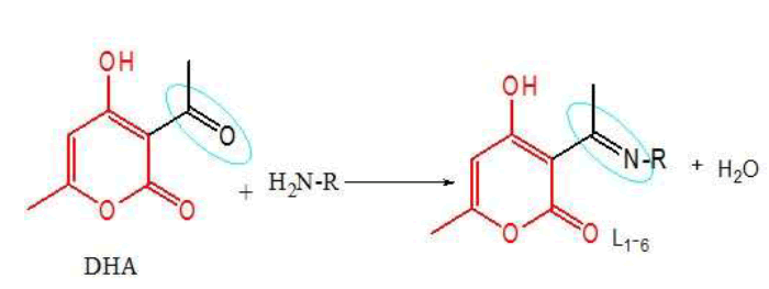
Schiff base motifs were synthesized by the addition of ethanolic solution of individual primary amine (0.05 mol) into hot ethanolic solution of DHA (3-acetyl-6-methyl (2H) pyran -2,4-(3H) dione) (0.05 mol) as per the procedure (Scheme 1) [10,11].
| Entries | R | Entries | R |
|---|---|---|---|
 |
|||
| L1 | 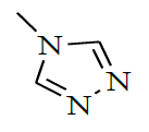 |
L4 | 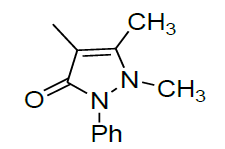 |
| L2 | 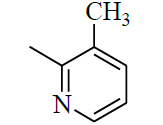 |
L5 | 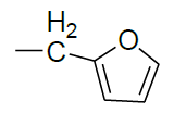 |
| L3 | 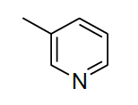 |
L6 | 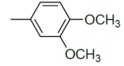 |
Scheme 1: A new series of compounds of dehydroacetic acid based Schiff bases derived from aromatic primary amines and synthesized
Results and Discussion
Elemental analysis
All the synthesized Schiff bases are white to yellowish to orange coloured solids, stable to air and non-hygroscopic. They are insoluble in water and soluble in hot ethanol. Their physical characteristics and elemental analysis data are summarized in Table 1.
| Entries | Molecular formula | Colour | Mol. Wt. | Melting point (°C) | Found/(Calculated)% | ||
|---|---|---|---|---|---|---|---|
| C | H | N | |||||
| L1 | C10H10N4O3 | white | 234 | 120 | 51.23 (51.28) | 4.32 (4.27) | 23.45 (23.93) |
| L2 | C14H14N2O3 | Yellow | 258 | 134 | 65.2 (65.11) | 5.39 (5.43) | 10.80 (10.85) |
| L3 | C13H12N2O3 | Yellowish | 244 | 117 | 64.01 (63.93) | 4.92 (4.92) | 11.41 (11.47) |
| L4 | C19H19N3O4 | Yellow orange | 353 | 210 | 64.46 (64.58) | 5.25 (5.38) | 11.8 (11.89) |
| L5 | C13H13NO4 | White | 247 | 131 | 63.25 (63.16) | 5.30 (5.26) | 5.69 (5.66) |
| L6 | C16H17NO5 | White | 303 | 115 | 63.43 (63.37) | 5.62 (5.61) | 4.70 (4.62) |
Table 1: Elemental analysis of the synthesized Schiff base motifs
Spectral characterization
FTIR analysis
The IR spectral data of Schiff bases is used to discuss various peaks in the spectra with respect to vibrations caused by different functional groups. The absorption peak pattern in IR spectra exhibits complex nature due to various vibrational modes. However with limited objectives, only characteristic peaks which are specific to all Schiff bases related to enolic O-H, aromatic C=C, azomethine >C=N, aryl azomethine, lactone carbonyl C=O and enolic C-O/C=O of Schiff bases have been taken into consideration for characterization.
In all the synthesized Schiff bases the characteristic O-H stretching frequencies were observed as broad weak band at 3438-3402 cm-1 due to strong intermolecular hydrogen bonding between enolic O-H and N of azomethine group. Azomethine >C=N stretching frequency is dependent on its substituent and mostly causing resonance interaction and H-bonding. In the present work azomethine (>C=N-) depicted strong absorption stretching vibration band in the region 1664-1630 cm-1. The peak in the region 1704-1685 cm-1 is appeared due to >C=O lactone carbonyl stretching vibrations, peak in the region 1392-1352 cm-1 is appeared due to aryl/aliphatic (C-N) stretching vibrations and the peak in the region 1288-1236 cm-1 is appeared due to (C-O) enolic stretching vibrations (Table 2) [11,12].
| Compounds | (O-H) | (> C=O) | (> C=N) | (> C=C <) | (C-N) | (C-O) |
|---|---|---|---|---|---|---|
| L1C10H10N4O3 | 3427 | 1689 | 1646 | 1571 | 1358 | 1256 |
| L2C14H14N2O3 | 3402 | 1689 | 1630 | 1573 | 1370 | 1288 |
| L3C13H12N2O3 | 3429 | 1703 | 1658 | 1558 | 1357 | 1266 |
| L4C19H19N3O4 | 3419 | 1699 | 1657 | 1577 | 1392 | 1259 |
| L5C13H13NO4 | 3430 | 1704 | 1664 | 1564 | 1352 | 1245 |
| L6C16H17NO5 | 3238 | 1685 | 1639 | 1532 | 1388 | 1236 |
Table 2: Characteristic IR frequencies (cm-1) of Schiff bases
1H-NMR spectra
1H-NMR spectra of all the compounds were recorded in CDCl3 at room temperature. The following signals, chemical shift value δ(ppm) relative to TMS as internal standard were observed.
Singlet signal at δ value 2.1-2.28 of methyl group at C6, singlet signal at δ value 14.72-16.00 of enolic O-H group which is highly deshielded, singlet signal at δ value 5.00-5.94 of H atom at C5 and singlet signal at δ value 2.57-2.75 of methyl group attached to azomethine C atom of DHA moiety and different signals of amine moiety [12].
3-((E)-1-(4H-1,2,4-triazol-4-ylimino)ethyl)-4-hydroxy-6-methyl-2H-pyran-2-one (L1): 1H-NMR (500 MHz, CDCl3, δ, ppm): 2.28 (3H, s, C6–CH3), 15.85 (1H, s, O–H), 5.94 (1H, s, C5–H), 2.67 (3H, s, N=C-CH3, H bonded to ‘C’ azomethine) for DHA moiety, 7.27 (2H, s, C–H), for triazole moiety.
3-((E)-1-(3-methylpyridin-2-ylimino)ethyl)-4-hydroxy-6-methyl-2H-pyran-2-one (L2): 1H-NMR (400 MHz CDCl3, δ, ppm): 2.1 (3H, s, C6– CH3), 15.85 (1H, s, O–H), 5.9 (1H, s, C5–H), 2.6 (3H, s, N=C-CH3, H bonded to ‘C’ azomethine) for DHA moiety, 2.2 (3H,s,CH3–Ar. ring), 7.7 (1H, m, C6-H), 6.9-7.2 (2H, m, C4 & C5-H), for pyridine moiety.
4-hydroxy-6-methyl-3-((E)-1-(pyridin-3-ylimino)ethyl)-2H-pyran-2-one (L3): 1H-NMR (400 MHz CDCl3, δ, ppm): 2.2 (3H, s, C6–CH3), 16.00 (1H,s,O –H), 5.8 (1H, s, C5–H), 2.6 (3H, s, N=C-CH3, H bonded to ‘C’ azomethine) for DHA moiety, 8.5-8.9 (2H, m, Ar. C2 & C6 H), 7.4- 7.7 (2H, m, Ar. C4 & C5-H) for pyridine moiety.
(4E)-4-(1-(4-hydroxy-6-methyl-2-oxo-2H-pyran-3-yl)ethylideneamino)-1,2-dihydro-2,3-dimethyl-1-phenylpyrazol-5-one (L4): 1H-NMR (500 MHz CDCl3, δ, ppm): 2.2 (3H, s, C6–CH3), 15.54 (1H, s, O–H), 5.75 (1H, s, C5–H), 2.7 (3H, s, N=C-CH3, H bonded to ‘C’ azomethine) for DHA moiety, 2.15 (3H, s, ring. –CH3), 3.2 (3H, s, ring N-CH), 7.4-7.6 (5H, m, Ar.) antipyrine moiety.
3-((E)-1-((furan-2-yl)methylimino)ethyl)-4-hydroxy-6-methyl-2H-pyran-2-one (L5): 1H-NMR (300 MHz CDCl3, δ, ppm): 2.11 (3H, s, C6– CH3), 14.82 (1H, s, O–H), 5.7 (1H, s, C5–H), 2.75 (3H, s, N=C-CH3, H bonded to ‘C’ azomethine) for DHA moiety, 4.7 (2H, s, =N-CH2 to Ar.), 7.5 (1H, m, C5), 6.45 (2H, m, C3 & C4) furan moiety.
3-((E)-1-(3,4-dimethoxyphenylimino)ethyl)-4-hydroxy-6-methyl-2H-pyran-2-one (L6): 1H-NMR (400 MHz CDCl3, δ, ppm): 2.15 (3H, s, C6–CH3), 15.7 (1H, s, O–H), 5.78 (1H, s, C5–H), 2.6 (3H, s, N=C-CH3, H bonded to ‘C’ azomethine) for DHA moiety, 3.94 (6H, s, two Ar. OCH3), 7.3 (1H, s, C2 of Ar.), 6.65-6.95 (2H, m, C5 & C6) aniline moiety.
Screening for bioactivity
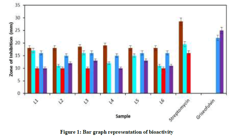
In vitro antibacterial and antifungal activities of the compounds were screened by considering zone of inhibition of growth. The synthesized compounds were screened with their different concentrations with standard antibiotics such as streptomycin (1 mg/ml) and griseofulvin (1 mg/ml) (Figure 1).
The compounds have shown harmonious antibacterial and antifungal action. All the compounds were found to be biologically active against E. coli with maximum zone of inhibition 19 mm by L1. Compound L1 has showed maximum zone of inhibition 17 mm against S. aureus. Compound L3 has showed 16 mm, L5 has showed 15 mm, zone of inhibition against S. aureus. The compounds L2, L4 and L6 were found to be moderate in growth inhibition against the same species. All the compounds were found to be moderate in growth with10 mm zone of inhibition while compound L4 and L5 are inactive against B. subtilis. All the compounds have shown harmonious antifungal action. All Compounds showed about 10-13 mm zone of inhibition and L3 and L5 depicted maximum zone of inhibition 13 mm against A. niger. All Compounds showed about 15-16 mm zone of inhibition against human opportunistic pathogen, C. albicans (Table 3).
| S. No. | Compound | Antibacterial (mm) | Antifungal (mm) | |||
|---|---|---|---|---|---|---|
| Escherichia coli | Staphylococcus aureus | Bacillus subtilis | Candida albicans | Aspergillus niger | ||
| 1 | L1 | 19 | 17 | 10 | 16 | 10 |
| 2 | L2 | 18 | 11 | 10 | 15 | 12 |
| 3 | L3 | 18.5 | 16 | 10 | 16 | 13 |
| 4 | L4 | 17 | 12 | 0 | 15 | 10 |
| 5 | L5 | 18 | 15 | 0 | 16 | 13 |
| 6 | L6 | 18 | 11 | 10 | 16 | 11 |
| 7 | Streptomycin | 28.5 | 19.5 | 16 | 0 | 0 |
| 8 | Griseofulvin | 0 | 0 | 0 | 22 | 25 |
Table 3: Summarized bioactivity data
Conclusion
All the Schiff bases are white to yellowish to orange coloured solids, stable to air and non-hygroscopic. The composition of all synthesized Schiff bases were confirmed by elemental analysis and their structures were determined by IR and 1H-NMR spectroscopic techniques. All the synthesized Schiff bases were found to possess strong antifungal activity and antibacterial activity. The compound L1 the Schiff Base of 4- amino-1,2,4-triazole exhibited the stronger inhibitor of growth against E. coli as compared to other Schiff bases. Least growth inhibitory activity was shown by compound L4 and L5 the Schiff bases of 4-amino antipyrine and 2-aminomethyl furan against B. subtilis species.
Acknowledgement
The authors express sincere thanks to UGC (WRO) for the award of Teacher Research Fellow (TRF) under UGC’s 11th plan period. Authors are also grateful to Principal R.S.M., Latur. Director, BCUD SRTM University, Nanded. Head of the Department of Chemistry & Analytical Chemistry and All the staff of Department of Chemistry & Analytical Chemistry. The authors also thankful to A. A. Yadav, V.D. Panchal, Reena Jadhav and special thanks to S.H. Kathwate, Department of Microbiology, Savitribai Phule Pune University and V.S. Shembekar Department of Biotechnology, Rajarshi Shahu Mahavidyalaya, (Autonomous) Latur.
References
- S. Kannan, R. Remesh, Polyhedron., 2006, 25, 3095.
- P.S. Mane, S.M. Salunke, B.S. More, T.K. Chondhekar, Asian J. Chem., 2012, 24(5), 2235.
- M.S. Karthikeyan, D.J. Prasad, B. Poojary, K.S. Bhat, B.S. Holla, N.S. Kumar, Bioorg. Med. Chem., 2006, 14, 7482.
- P. Vicini, A. Geronikaki, M. Incerti, B. Busoners, G. Poni, C.A. Cabras, P.L. Colla, BBioorg. Med. Chem, 2003, 2, 4785.
- S.M. Jadhav, V.A. Shelke, S.G. Shankarwar, A.S. Munde, T.K. Chondhekar, J. Saud. Chem. Soc., 2014, 14, 27.
- A.M. Khedr, M. Gaber, R.M. Issa, H. Erten, Dyes Pigm., 2005, 67, 117.
- R.M. Issa, A.M. Khedr, H. Rizk, J. Chin. Chem. Soc., 2008, 35(4), 875-884.
- A.I Vogel, Text Book of Practical Organic Chemistry, 5th Edi., Longman, London, 1989, 400-401.
- W. Rehman, Z. Hassan, U. Rashid, F. Rahim, O.U. Rehaman Abid, M. Waseem, Med. Chem. Res., 2014, 23, 2307-2311.
- R.G. Bor, 1913, 46, 2712.
- D.G. Palke, S.D. Salunke, Asian J. Chem., 2017, 29, 2167-2170.
- Spectroscopic Identification of Organic compounds, Silverstein, Bassler and Morril, 4th Edi, John Wiley and Sons New York, 1980, 196, 226, 230.

