Research Article - Der Pharma Chemica ( 2021) Volume 13, Issue 3
Synthesis, characterization and antibacterial activity of Schiff base of Cu (II), Ni (II) and Co (II) complexes
Salah Hamza Sherif*, Dagne Addisu Kure1, Endalkachew Asefa Moges and AkmalNur Negash2Department of Anthropology, Hawassa University, Hawassa, Ethiopia
Salah Hamza Sherif, Department of Chemistry, Hawassa University, Hawassa, Ethiopia, Email: hamzasalahsherif@gmail.com
Abstract
Due to their importance as catalysts in many reactions and their biological activities, an interest in the synthesis and characterization of transition metal complexes containing Schiff bases is increasing. The major aim of this study was to compare the antibacterial activity of the ligands with their metal complexes. We have successfully synthesized Cu (II), Ni (II) and Co (II) complexes of N, N-Bis(vanillinidene )-1,2-phenylenediamine and N,N-Bis(salicylidene)-1,2-phenylenediamine. The Schiff’s bases were synthesized by the reaction of o-phenylenediamine with salisaldehyde or o-vanillin and their metal complex were synthesized by the reaction of the ligands with the metal salts. The structure of all the synthesized ligands were confirmed using NMR, IR and UV-Vis spectral analysis and their metal complexes were confirmed using IR and UV-vis spectra. The synthesized ligands and their metal complexes were screened for their antibacterial activities against Escherichia coli, Staphylococcus aureus, and Klebsiella pneumonia bacterial strains by using disc diffusion method. Among the synthesized compounds, the ligand L1 is moderately active against Staphylococcus aureus, K. pneumonia and Escherichia coli bacteria strain, CoL1 exhibited the highest activity, with 22mm zone of inhibition against staphylococcus aureus, compared to standard gentamycin activity, with 21 mm zone of inhibition.
Keywords
o-phenylenediamine; o-vanillin; Salisaldhyde; Schiff base; Transition metal (II) complexes; Antibacterial activity
Introduction
Schiff’s base ligands are usually prepared by condensation between aldehydes and primary amines. They are the most widely studied ligands and are considered as ‘‘privileged ligands’’ [1,2] and are able to coordinate many different metals to stabilize them in various oxidation states [3].
Multidentate Schiff bases have been widely used as ligands, because they can be easily attached to metal ions to generate stable complexes. It interacts with most metallic ions and especially with transition ones and have ability to stabilize them in various oxidation states. Schiff bases have played an important role as chelating ligands for a large variety of metal ions.Recently, there has been enhanced interest in the synthesis and characterization of transition metal complexes containing Schiff bases due to their importance as catalysts in many reactions [4-10].
Transitionmetal complexes of Schiff’s bases particularly derived from carbonyl compounds based on heterocyclic rings have also received great attention in medicinal and pharmaceutical field, a number of compounds containing Schiff base have been synthesized and tested for their biological activity, because they exhibited broad range of biological activities, such as antibacterial [11-13],anticancer [14-18], anti-inflammatory [19],antioxidant [20-22] etc.
In this paper, we present the synthesis and anti-bacterial activity investigations of N, N-Bis(vanillinidene )-1,2-phenylenediamine and N, N-Bis(salicylidene)-1,2-phenylenediamine and their metal complex against K. pneumoniae, E. coli, and S. aureus bacterial strain. The major aim of this study was to compare the activity of the ligands with their metal complexes.
Experimental Section
Materials and methods
All chemicals and solvents were analytical grade and used without further purification; the progress of the reaction was monitored by TLC silica gel plates; the purification of the products was performed by column chromatography using silica gel (100–200mesh). Melting points were measured in open capillary tubes and were uncorrected; infrared (IR) spectra were recorded using FT-IR Bruker Alpha spectrometer.NMR spectra were recorded on BrukerAvance NMR spectrometer (400MHz) using TMS as the internal standard.
Chemistry
The free ligands and their metal complexes were prepared as outlined in Scheme 1. The Schiff’s bases were synthesized by the reaction of o-phenylenediamine with salisaldehyde or o-vanillin. The product was further purified by column chromatography using mixture of hexane and ethyl acetate as solvents system in different ratio. The ligands were further reacted with copper (II) acetate, nickel (II) acetate and hydrated cobalt (II) chloride. The mixture was refluxed at a temperature of 70-80 °C and the progress of the reaction was monitored by thin layer chromatography (TLC). The solid product formed was separated by filtration, washed with ethanol and recrystallized from chloroform and dried.
Synthesis and characterization
General procedure for the Synthesis of N, N-Bis (vanillinidene)-benzene-1, 2-diamine (L1) and N, N-Bis (salicylidene)-benzene-1, 2-diamine (L2)
O-phenylenediamine (0.054g, 0.5mmol) was dissolved in 10ml ethanol and a solution of o-vanillin (0.304g, 1mmol) or salisaldehyde (0.244g, 1 mmol) in 20ml ethanol was added to this mixture, 3-5 drops of glacial acetic acid was added and refluxed at 70-80°C for 2 hours.The progress of the reaction was monitored by using TLC. After completion of the reaction, the reaction mixture was cooled and ethanol was removed using rotary evaporator.The solid obtained was further purified by column chromatography using mixture of n-hexane: ethyl acetate solvent system.
Characterization of L1
Reddish yellow solid, Yield: 75 %, Mp: 161-163°C; 1H NMR (DMSO-d6, 400MHz): δH 13.20(2H, OH), 7.57 (s, 2H, azomethine), 6.69-7.09(m, 10H, aromatic proton), 3.73(s, 6H, methoxy proton). 13C NMR (DMSO-d6, 100MHz): δC 164.1, 152.1, 151.0, 142.5, 128.3, 124.9, 123.7, 122.9, 119.6, 118.7, 56.2
Characterization of L2
Orange solid, Yield: 68.1 %, Mp: 206-208 °C; IR(KBr, Cm-1): 3394(-NH), 3240(-OH), 3047(C-H, aromatic), 2854(C-H, methyl), 1593(C=N);1H NMR (DMSO-d6,400MHz): δH 12.9(s, 2H, OH). 8.1(s, 2H, azomethine proton) 6.96-7.4(m, 12H, aromatic proton); 13C NMR (DMSO-d6, 100MHz): δC 161.2, 160.3, 142.7, 132.5, 130.6, 128.8,1 23, 121.5, 118.5, 116
Synthesis of Cu (II), Ni (II) and Co (II) complexes of ligand L1
The ligand L1 (0.224g, 0.5mmol) was dissolved in 10ml of hot ethanol, and mixed with (0.5mmol) of Cu(CH3COO)2.H2O,Ni(CH3COO)2.4H2O or CuCl2.6H2O dissolved in 5ml of hot ethanol. The reaction mixtures were refluxed for 1 hour. The progress of the reaction was monitored using TLC. After the reaction was completed, the reaction product was cooled in ice water, filtered and washed with ethanol. Finally, the solid was recrystallized from chloroform and dried. The physical characteristics and molar conductance of L1, L2 and their metal (II)complexes given in Table 1.
| No | Compound | Color | % yield | Molar conductance(μs/cm) |
|---|---|---|---|---|
| 1 | L1 | Reddishyellow | 75 | 27.1 |
| 2 | L2 | Orange | 68.1 | 25.6 |
| 3 | Cu (II)L1 | Yellowish brown | 79.64 | 24.6 |
| 4 | Ni (II)L1 | Black red | 74 | 18.8 |
| 5 | Co (II)L1 | Brown | 42.13 | 22.4 |
| 6 | Cu (II)L2 | White brown | 59.89 | 20.5 |
| 7 | Ni (II)L2 | Black red | 80.6 | 14.14 |
Synthesis of Cu (II), Ni (II) and Co (II) complexes of ligand L2
The ligand L2 (0.16 g, 0.5 mmol) was dissolved in 10 ml of hot ethanol, and mixed with (0.5mmol) of Cu(CH3COO)2.H2O, or Ni(CH3COO)2. 4H2O dissolved in 5 ml of hot ethanol. The reaction mixtures were refluxed for 1 hour. The progress of the reaction was monitored using TLC. After the reaction was completed, the reaction product was cooled in ice water, filtered and washed with ethanol. Finally, the solid was recrystallized from chloroform and dried.
The Uv-visble spectra of the ligand, L1 and its complexes were recorded in DMSO at room temperature .The observed spectrum of ligand L1 showed three intense bands around 278 nm, which was assigned to π→π* transition of C=N, 291nm assigned to n→π* transition of C-O methoxy group, 300nm transition of the OH. In complex formation this was shifted to a higher wavelength suggested the coordination of azomethine nitrogen. The UV-Vis spectra of copper complex showed three bands at 301nm assigned to π→π* transition from C=N, 382 nm assigned to n→π* transition from the azomethine nitrogen of ligand to metal charge transfer and 493nm n→π* transition from hydroxyl oxygen of ligand to metal charge transfer. Nickel (II) complexes showed three absorption bands at 301nm π→π* transition of C=N, 382nm n→π* transition from azomethine nitrogen of the ligand to metal charge transfer, 496nm n→π* transition from the hydroxyl of the ligand to metal charge transfer. Cobalt(II) complex of L1 showed three absorption bands at 301nm π→π* transition of azomethine C=N group, 381nm transition from the azomethine nitrogen of the ligand to metal charge transfer, 496nm transition fromthehydroxyl oxygen of the ligand to metal charge transfer.
The UV-visible spectra of the ligand, L2 and its metal complexes were recorded in DMSO-d6 at room temperature. The electronic spectra of ligand L2 showed two bands at 273nm which is assigned to π→π* transition of C=N group, 333nm n→π* transition from OH. UV of Cu (II) complex showed three bands at 301nm π→π* transition of azomethine group(C=N), 381nm n→π* transition from azomethine nitrogen of the ligand to metal charge transfer and 493nm n→π* transition from the hydroxyl oxygen of the ligand to metal charge transfer. The Ni (II) complex exhibit three bands at 300nm π→π* transition of the azomethine C=N group, 382nm n→π* transition from azomethine nitrogen of the ligand to metal charge transfer and 495nm n→π* transition from the hydroxyl oxygen of the ligand to metal charge transfer.
Results and Discussions
Antibacterial activities
Culture media and disk preparation
Nutrient agar, Muller Hinton agar and Nutrient broth were prepared according to themanufacturer instruction in which the prepared media was autoclaved at 121°C for 15minutes. Then the prepared culture media was checked for the sterility for 24 hours at 37°C. Quality control stains of Staphylococcus aureus, Escherichia coli and Klebsiella pneumonia known. American type culture collection committee (ATCC) was used to perform theantibacterial activities of the agents. Watman filter paper was used to prepare a disk of 5mm diameter using manual paper punching.
Preparation of chemical solution and media for the antibacterial activity
By using analytical balance, a 500 μ g of each chemical powder was added to 15ml dimethyl sulfoxide (DMSO) and mixed to form a homogenous solution. A 5μl of solution was added to the sterile disk prepared before using sterile micropipette. Quality control strains obtained from Hawassa University College of medicine and health science was inoculated on nutrient agar plate using sterile loop (Table 2).
| Compound (500μg/5μL disc) | Inhibition zone(mm) | ||
|---|---|---|---|
| Staphylococcus aureus | Klebsiella pneumonia | Escherichia coli | |
| L1 | 13 | 7 | 9 |
| CuL1 | 7 | 5 | 5 |
| Ni L1 | 10 | 5 | 11 |
| Co L1 | 22 | 5 | 5 |
| L2 | 11 | 5 | 5 |
| CuL2 | 7 | 5 | 5 |
| NiL2 | 5 | 5 | 5 |
| Gentamicin | 21 | 6 | 21 |
The plate was incubated for 24 hours at 37 °C. A 3-5 colonies was picked and suspended in 5ml nutrient broth to form agar plate in three directions to form uniform inoculums, then a disk with a control gentamicin and solution impregnated disk was placed on the plate and incubated for 24 hours at 37 °C. Each disk was labeled with its unique ID number on the back of the Petri dish. After incubation the diameter of the zone of inhibition was measured using ruler. Growth of bacteria pathogens on each concentration was checked to determine the minimum concentration that inhibits the growth of bacteria’s. It is evident from table 2 that the minimum concentration value for each ligands and its metal complexes were (500 μ g/5 μ L disc) against Staphylococcus aureus, Klebsiella pneumonia and Escherichia coli.
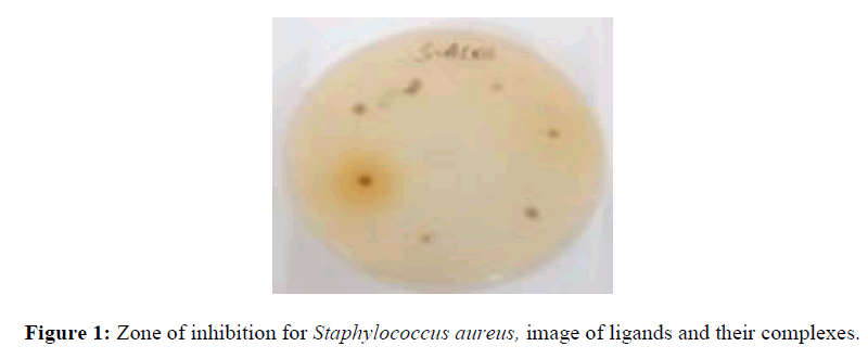
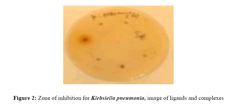
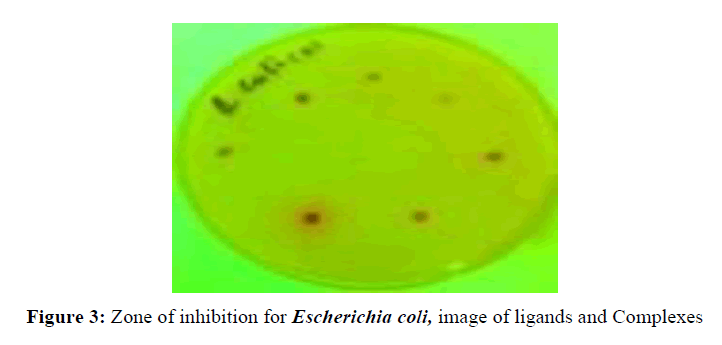
The antibacterial activity of the ligands and complexes were measured based on zone of inhibition on the grown bacteria on the prepared culture media on petri-dish as shown (Figures 1-3) its inhibition value were measured using ruler.
Conclusion
The antibacterial activity of the ligands and their metal complexes were studied against three pathogenic bacterial strains, one gram positive (Staphylococcus aureus) and two gram negative (Klebsiella pneumonia and Escherichia coli) Table 2. The ligand L1 is moderately active against Staphylococcus aureus, K. pneumonia and Escherichia coli bacteria strain, whereas NiL1 complex exhibited less activity against those bacteria strain and CoL1 exhibited the highest activity against Staphylococcus aureus. CuL2 and NiL2 exhibited no significant activity against all the tested bacterial strain. In general, the ligand L1 and its metal complexes have higher zone of inhibition than L2 and its metal complexes. Comparing the two ligands, L1 showed better activity than L2, structurally the two ligands differ by the presence of methoxy substituent, the difference in activity may be due to this substituent.
Acknowledgments
Authors are thankful to Hawassa University for providing research fund, Hawassa University College of medicine and health science for antibacterial screening and Addis Ababa University for running NMR spectra.
References
- Singh KB, Prakash A, Rajour KH et al. Spectrochim. Acta, 2010.76: p. 376–383.
- Abdel-Latif AS, Hassib BH and Issa MY. Spetrochimica Acta A. 2007. 67: p. 950.
- HosseinNaeimi and Mohsen Moradian., J. Chemistry. 2013.
- Gaurav B Pethe, Amit R Yaul and Anand SAswar, Bull. Chem. Soc. Ethiop. 2015. 29: p. 387-397.
- Seitz M, Kaiser A,Stempfhuber S et al., J. Amer. Chem. Soc, 2004. 126: p. 11426–11427.
- Singh DP, Kumar R, Mehani R et al., J Serb ChemSoci, 2006. 71: p. 939.
- Deepa NT, Madhu PK, Radha Krishnan. Synth. React. Inorg Met.Org. chem., 2005.35: p. 883-888.
- Z. H. Chohan, H. Pervez, A. Rauf, K. M. Khan, C. T. Supwern,J. Enzyme Inhib. Med. Chem, 2002, 17, 117–122.
- Ali SA, Soliman AA, Aboaly MM et al., J Coord Chem, 2002. 55: p. 1161-1170.
- Chatterjee D, Mitra A and Roy BC. J. Mol. Cat, 2000. 16: p. 17-21.
- ShridharMalladi, Arun M. Isloor, Shrikrishna Isloor et al., Arab J Chem, 2013. 6: p. 335–340.
- Ikechukwu P. Ejidike and Peter A. Ajibade, Molecules, 2015. 20: p. 9788-9802.
- Navin B. Patel, Jaymin C. Patel, Arab J Chem, 2011. 4: p. 403–411.
- Sanaa M. Emam, Ibrahim ET El Sayed, Mohamed IAyad et al., J MolStruc, 2017.1146: p. 600-619.
- Mokhles M. Abd-Elzaher , Ammar A. Labib, Hanan A. Mousa et al., J basic app scien, 2016. 5: p. 85–96.
- SubinKuamr K, PriyaVarma C, VN Reena et al., J. Pharm. Sci. & Res. 2017, 9: p. 1317-1323.
- ShanshanShen, Hong Chen, Taofeng Zhu et al., Oncology Letters, 2017. 13: p. 3169-3176.
- Deodware SA, Sathe DJ, Choudhari PB et al., Arab J Chem, 2017. 10: p. 262–272.
- Nasser Mohammed Hosny , Yousery E. Sherif&Amany A. El-Rahman, J CoordChem, 2008. 61: p. 2536–2548.
- Saif M, HodaF.El-Shafiy, Mahmoud M. Mashaly et al., J MolStruc, 2016. 1118: p. 75-82.
- Mruthyunjayaswamy BHM, Nagesh GY, Ramesh M et al., Der PharmaChemica, 2015. 7: p. 556-562.
- Desireddy Harikishore Kumar Reddy, Seung-Mok Lee1, Kalluru Seshaiah et al., J. Serb. Chem. Soc., 2012. 77: p. 229–240.


