Research - Der Pharma Chemica ( 2021) Volume 13, Issue 9
Protective Effect of Brassica olearacea Var. Capitata Extract on Glutathione-s-transferase, Catalase and Total Protein Activity in Dolutegravir-induced Toxicity
Amagon L*, Falang KD, Bukar BB, Wanche EM and Amagon KIAmagon L, Department of Pharmacology & Toxicology, Faculty of Pharmaceutical Sciences, University of Jos, Jos, Nigeria, Tel: +234-803-3713242, Email: leritshimwa@yahoo.co.uk
Received: 18-Aug-2021 Accepted Date: Sep 24, 2021 ; Published: 30-Sep-2021
Abstract
Introduction: Oxidative stress is one of the leading causes of liver injury; the process of such damage involves the molecular reactions of highly reactive oxygen species that can lead to impaired liver functions.
Objective: This study investigated the protective effect of Brassica oleracea (Red Cabbage against Dolutegravir-induced oxidative stress and assessed the safety of the plant extract in Drosophila melanogaster.
Methods: In the first phase, there were three groups of flies in replicates of five (5), with each vial contained 50 flies of both gender; group one was the control, and the other flies were administered the toxicant for ten days, then homogenized, and the biochemical assays performed. In the second phase, there were five groups of flies in replicates of five (5), each vial containing 50 flies. They were administered the extract for seven days, exposed to the toxicant for Five (5) days, and homogenized. The biochemical assay was carried out to determine the level of glutathione-s-transferase and catalase.
Results: Brassica oleracea extract was observed to be safe in the flies exposed to the extract. Glutathione-s-transferase and catalase activity was reduced in the group treated with dolutegravir but increased in the presence of different concentrations of Brassica oleracea.
Conclusion: The extract of Brassica oleracea showed a good ability to protect against dolutegravir-induced toxicity and was also safe in D. melanogaster.
Keywords
Brassica oleracea, Diet, Dolutegravir, Oxidative stress, Total thiol
Introduction
Oxidative stress results from an imbalance between the free radicals present in the body and the antioxidant activity in the body. It arises when the free radicals exceed the antioxidants in the body. Unpaired electrons form pairs with other electrons, thus making free radicals unstable and quite reactive.
For aerobic organisms, a mechanism to remove these highly reactive oxygen species is essential to sustain life. Diseases can be caused by free radicals and oxygen species that attack cellular membranes and tissues. Fruits high in vitamin C are taken to clean out the excess free radicals and restore a balance between the free radicals and the antioxidants.
Oxidative stress has been implicated in the pathophysiology of some diseases such as cardiovascular insufficiency, and vascular stenosis. It is a leading cause of liver injury, and the process involves the molecular reactions of highly reactive oxygen species in the liver, which can lead to impaired liver functions and other systemic complications [1].
Biomarkers that can be used to assess oxidative stress have attracted interest because a right assessment is necessary for investigating various pathological conditions [2]. Superoxide dismutase, total thiols, glutathione-s-transferase, catalase, acetylcholinesterase, and glucose-6-phosphate Dehydrogenase (G6PDH) are biomarkers of oxidative stress in fruit flies [3].
Dolutegravir, an HIV integrase inhibitor, acts by impairing the function of the HIV integrase-DNA complex to which it was synthesized to bind. Urinary excretion is minimal, as it is predominantly metabolized via hepatic glucuronidation by UDP-glucuronosyltransferase [4]. Reported side effects include increased creatinine kinase, increased aspartate aminotransferase (AST), Insomnia, increased alanine aminotransferase (ALT), increased bilirubin, increased cholesterol and triglycerides, increased lipase, and hyperglycemia [5-7].
Brassica oleracea Var Capitata is an herbaceous biennial plant of the family Brassicacea that forms a compact head. It is also known as red cabbage and is a good source of iron, vitamin A and vitamin C, and is capable of boosting the body’s immunity against diseases [8]. This plant contains powerful antioxidants that may help reduce inflammation [9]. In a previous study among several species of Brassicacea grown in Malaysia, red cabbage showed the highest antioxidant activity and phenolic content, compared to the other vegetables studied [10].
Rathore and colleagues [11] concluded in their review, that the potent hepatoprotective activities of chemically defined molecules isolated from plants signify a stimulating lead in the search for effective and inexpensive liver-protecting agents. Their findings show that leaf extracts and extracts from other plant parts have good promise for use in liver disease and may offer new possibilities to the limited therapeutic options that presently exist in the treatment of liver diseases, and should be considered for future research.
Drosophila is a genus of small flies, belonging to the family Drosophilidae, whose members are known as “fruit flies”. Drosophila melanogaster, a species of Drosophila, has been used as an animal model in genetic research and developmental biology. Most organisms share basic genetic mechanisms, and this is vital as many Drosophila genes are homologous to human genes. These are studied to gain a better understanding of what role these proteins play in humans.
Materials and Methods
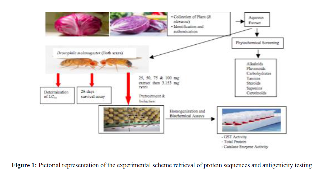
Collection and Identification of Plant: The plant was purchased at Farin Gada market in Jos North Local Government Area, Jos Plateau State. Mr. Joseph Azila, a taxonomist at the Federal College of Forestry, Jos, Nigeria, authenticated and identified the plant material. A sample was deposited at the College herbarium with specimen number FHJ 33221.
Extraction: The leaves of the red cabbage were removed, sliced into pieces using a knife, and then blended with an electric blender. About 500 ml of water was added to 500 g of the blended sample and allowed to stand overnight in a refrigerator. The sample was filtered using a sieve and the filtrate was concentrated to dryness using a water bath at 70 °C and concentrated in a drying cabinet. The extract collected was weighed to be preserved in a refrigerator in a tight stride sample container till required for use.
Phytochemical Screening
Phytochemical analysis was conducted to determine the metabolites present in the extract. The presence of flavonoids, terpenoids, saponins, alkaloids, glycosides, carbohydrates, and proteins was determined using various methods.
Experimental Design
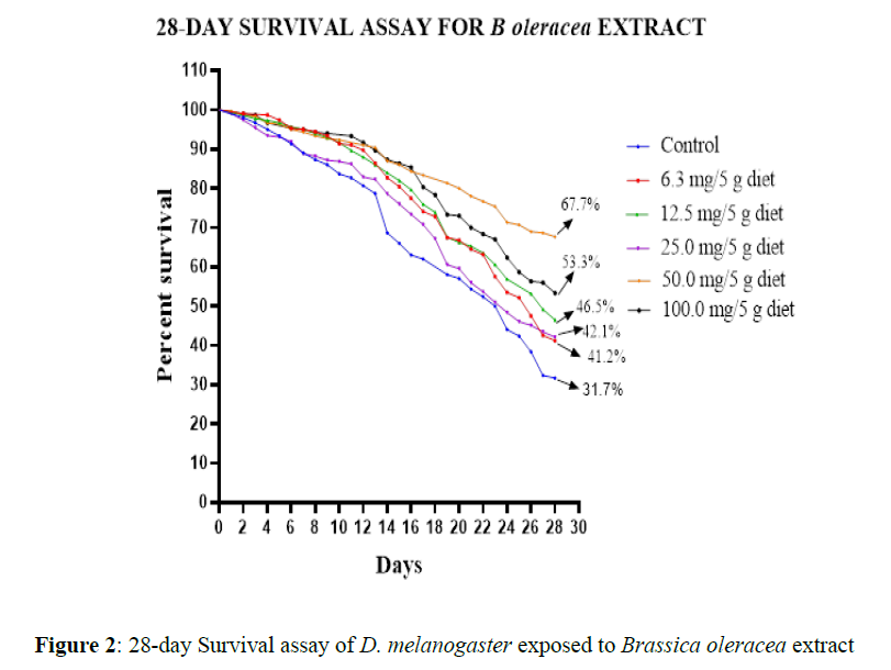
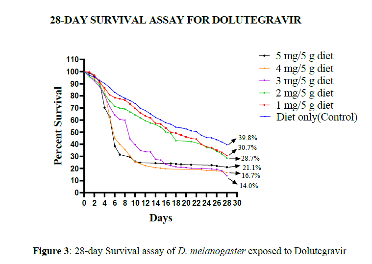
A pilot assay was done on Drosophila melanogaster to determine a suitable concentration that would not result in outright mortality. This was done by exposing the flies to various concentrations of DTG and the plant extract for 7 days, after which the survival assay was done by exposing the flies to various concentrations of the LC50 for 28 days and the number of dead and living flies was recorded daily.
Induction of Toxicity and Treatment
Phase One (Baseline): Three (3) groups of flies were obtained, each group contained a replicate of five (5) vials of 50 flies each of both gender. The first group was the control group and the flies were administered the normal diet for 10 days, after which the flies were homogenized and the biochemical assays were performed. The second and the third group had flies that were administered the LC30 of the toxicant (DTG). After 10 days, the flies were homogenized and the biochemical assays were conducted.
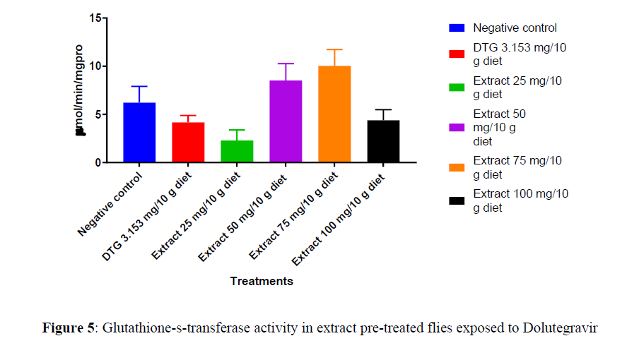
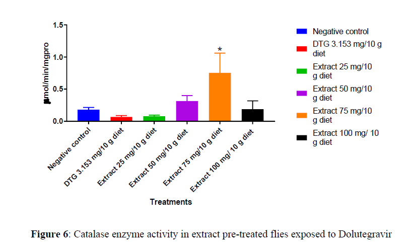
Phase Two (Preventive): Five groups of the flies were obtained, each group contained a replica of five (5) vials of 50 flies each of both gender. The first group was the control group and the flies were administered the normal diet for 10 days, after which the flies were homogenized and the necessary biochemical assays were conducted. The second, third, fourth, and fifth groups were administered graded concentrations (25 mg, 50 mg, 75 mg, and 100 mg) of the plant extract for 7 days, after which they were administered the toxicant for 5 days. The flies were homogenized, and the assays were conducted (Figures 1-6).
Statistical Analysis: One-way ANOVA and Tukey’s multiple comparison tests were used to analyze the data with the aid of GraphPad Prism 5 (GraphPad Software Inc., CA). (Table 1)
| Phytochemical | Remark |
|---|---|
| Alkaloid | +++ |
| Saponins | --- |
| Carbohydrates | +++ |
| Tannins/phenols | ++ |
| Flavonoid | +++ |
| Steroids | + |
| Terpenoids | --- |
| Cardiac glycosides | ++ |
Key: + Positive; - Negative
Results
There was a significant difference (p<0.05) comparing the survival curves, at 25 mg and 75 mg /10 g, survival proportion significantly increased (p<0.05) compared to control with a significant decrease (p<0.05) in survival proportion at 50 mg and 100 mg/10 g diet.
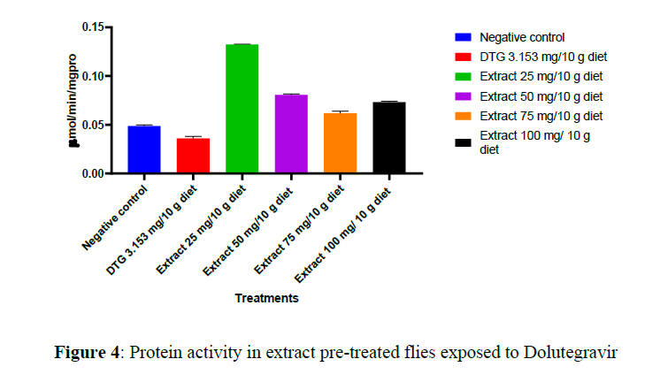
There was an elevation of GST activity in 50 mg, 75 mg, and 100 mg /10 g diet but no significant difference (p>0.05) compared to the DTG-treated group.
There was an elevation of catalase activity in 50 mg and 75 mg/10 g diet but only 75 mg/10 g diet significantly (p<0.05) elevated this activity compared to the DTG-treated group.
Discussion
Endogenous antioxidants exist to fight the free radicals in the body, in addition to the enzymatic antioxidants. These include glutathione, bilirubin, total proteins, catalase, superoxide dismutase, albumin, and uric acid, which as a whole play a homeostatic role against ROS produced during normal cellular activity [12].
Drosophila melanogaster is a suitable organism for toxicological studies because of its inexpensive maintenance, short generation time, high productiveness, comparatively short life, and well-characterized genome [13]. Other features that support the fruit fly's suitability as an experimental model are an intestinal system that looks like that of mammals, as well as a fat body that functions like the mammalian liver.
The phytochemical analysis of the plant extract in this present study revealed the presence of alkaloids, carbohydrates, tannins/phenol, flavonoids, steroid, and cardiac glycosides. The presence of alkaloids, tannins, phenols, and flavonoids imparts the plant its antioxidant activity [14]. The finding from this present study is similar to that from an earlier study that identified glycosides, flavonoids, triterpenes, and phenolic compounds as classes of compounds with hepatoprotective activity [11].
Dolutegravir, an HIV integrase inhibitor, has been reported to generate reactive oxygen species, leading to oxidative stress [15]. In this present study, the 28-day survival assay showed a dose-dependent decrease in the survival for fruit flies exposed to dolutegravir in their diet, compared to the flies that were administered a toxicant-free diet.
In this present study, the fruit flies would be expected to benefit from the presence of flavonoids and other phytochemicals in Brassica oleracea, which were incorporated in their diet throughout this study. This point is buttressed by results from this present study where the fruit flies exposed to the extract in their diet survived better compared to those in the control who fed on an extract-free diet. Dietary phytochemicals like flavonoids have reported good antioxidant activity [16].
Catalase is an important antioxidant enzyme responsible for the degradation of reactive oxygen species. It is widely expressed in both mammalian and non-mammalian cells containing the cytochrome system [17]. Reduction in the activity of the enzyme will lead to a high amount of circulatory ROS/RNS, as seen in this present study where a reduction in catalase was observed in flies exposed to dolutegravir, a drug known to generate reactive species. It has been suggested that the increase in antioxidant enzymes may represent a compensatory upregulation in response to increased oxidative stress [18,19].
Glutathione S-transferase (GST) plays a vital role against cancer-causing agents, drugs, and kinds of oxidative damage [20]. The biochemical assay in the fruit flies exposed to the toxicant showed a reduction in the level of Glutathione-s-transferase (GST), indicating induction of oxidative stress. The extract in this present study was able to offer protection, which was seen by an increase in levels of this antioxidant enzyme.
Reactive Oxygen species are very sensitive, with a short half-life, thus making measuring them in vivo to determine oxidative stress difficult [21]. In its place, lipids, proteins, and carbohydrates, which have much longer half-lives after being affected by ROS, are used as biomarkers of oxidative stress. In this present study, the protein level was observed to decrease following administration of dolutegravir in the diet of the fruit flies, compared to flies administered the diet alone.
Conclusion
The aqueous extract of Brassica oleracea protected against dolutegravir-induced oxidative stress in Drosophila melanogaster.
References
- Ahmed Z, Ahmed U, Walayat S et al., Clin Exp Gastroenterol., 2018, 11: p. 301-307.
- Beckman JA, Paneni F, Cosentino F et al., Eur Heart J. 2013, 34(31): p. 2444-2452.
- Lushchak OV, Rovenko BM, Gospodaryov DV et al., Comp Biochem Physiol A Mol Integr Physiol. 2011, 160: p. 27-34.
- Kandel CE and Walmsley SL. Drug Des Devel Ther. 2015, 9: p. 3547-3555.
- van Lunzen J, Maggiolo F, Arribas JR et al., Lancet Infect Dis. 2012, 12(2): p. 111-118.
- Walmsley SL, Antela A, Clumeck N et al., N Engl J Med. 2013, 369(19): p. 1807-1818.
- Blake M and Sonia V. Future Virol. 2015, 9(11): p. 967-978.
- Nolte E, Conklin A, Adams J et al., Santa Monica (CA)/Cambridge: RAND Corporation; 2012.
- Cruz MP, Andrade CMF, Silva KO et al., PLoS ONE. 2016, 11(3): p. e0150839.
- Lee WY, Emmy H, Khairul I et al., Mal Journal of Nutrition. 2007, 13(1): p. 71-80.
- Rathore PS, Rao P, Roy A et al., Research J Pharm and Tech. 2004, 7(2): p. 229-234.
- Vendemiale G, Grattagliano I, Altomare E et al., Ind J Clin Lab Res. 1999, 29: p. 49–55.
- Baenas N and Wagner AE. Genes Nutr. 2019, 14: p. 14.
- Selamoglu Z, Dusgun C, Akgul H et al., Iran J Pharm Res. 2017, 16: p. 92-98.
- Iorjiim WM, Omale S, Bagu GD et al., J Adv Med Pharm Sci. 2020, 22(6): p. 26-40.
- Knekt P, Kumpulainen J, Järvinen R et al., Am J Clin Nutr. 2002, 76(3): p. 560-568.
- Gutteridge JMC and Halliwell B. Trends Biochem Sci. 1990, 15: p. 129-130.
- Turk HM, Sevinc A, Camci C et al. Asian Pac J Cancer Prev. 2002, 39(3): p. 117-122.
- Likidlilid A, Patchanans N, Peerapatdit T et al., J Med Assoc Thai. 2010, 93(6): p. 682-693.
- Hayes JD, Flanagan JU and Jowsey IR. Annu Rev Pharmacol Toxicol. 2005, 45(1): p. 51-88.
- Pryor WA. Annu Rev Physiol. 1986, 48: p. 657-667.

