Full Length Research Article - Der Pharma Chemica ( 2022) Volume 13, Issue 8
Green Synthesis of Silver Nanoparticles Using Polyphenolic Rich Root Extract of Codonopsis Clematidea and Evaluation of Their Biological Potential
Ajay Sharma1*, Sarvpreet Singh1, Love1 and Pushpender Bhardwaj22Department of Medicinal Plant, Defence Institute of High-Altitude Research, Gharuan, India
Ajay Sharma, Department of Chemistry, University Institute of Sciences, Gharuan, India, Email: sharmaajay9981@gmail.com
Received: 09-Sep-2019, Manuscript No. dpc-20-2291; Editor assigned: 13-Sep-2019, Pre QC No. dpc-20-2291; Reviewed: 27-Sep-2019, QC No. dpc- 20-2291; Revised: 20-Jul-2022, Manuscript No. dpc-20-2291; Published: 17-Aug-2022, DOI: 10.4172/0975-413X.16.1.232-238
Abstract
Plants have wide range of medicinal values and have been used from ancient time to cure various kind of illnesses. India comes under the twelve mega-biodiversity countries of the world, which are rich in vegetation and have a wide variety of medicinal plants. From dawn of civilization, folks in India and China used medicinal plants to heal their wounds and various kinds of sicknesses. Codonopsis is a continual herb belongs to family Campanulaceae. Different studies depicted this plant as an important herb for various therapies. It is necessary to study this plant more broadly to know its medicinal values and use in the treatment of various diseases. Keeping this in view, in the present work green synthesis of silver nanoparticles using polyphenolic rich root extract of Codonopsis clematidea has been carried out. Characterization of synthesised nanoparticles has been carried out using UV-Visible, IR, XRD and TEM. The synthesized silver nanoparticles have also been evaluated for their biological potential.
Keywords
Codonopsis clematidea; Polyphenolics; Silver nanoparticles; Biological potential
INTRODUCTION
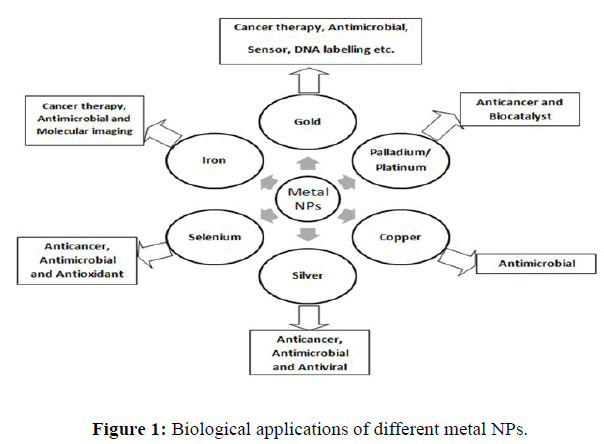
Metal NPs synthesized by green methods have attained world-wide consideration because of their unique physicochemical properties and their applications in the various fields of science [1]. In current years, the growth of synthesizing NPs by different plant extracts has become a key focus of researchers from all around the world because these NPs are eco-friendly and less toxic for the human body. The metal NPs synthesized using plants are not only more stable in terms of shape and size, but also the percentage yield of this technique is higher than the other approaches. These metal NPs have well known for their biological potential such as antimicrobial, antifungal, antioxidant, anticancer etc. [2]. Plant extracts serve two function, these have been act as reducing agent as well as stabilizer of metal NPs [3]. The plant extracts are rich in various Secondary Metabolites (SMs) such as polyphenols, flavonoids, steroids, glycosides, amino acids, sugars, tannins, lignins etc. These SMs act as reducing agents during green synthesis of metal NPs and are extremely helpful for the human body due to their wide range of biological potential such as anticancer, antiinflammatory, insecticidal, antioxidant, antimicrobial, anti-diabetic etc.[4]. Figure 1 presents the biological applications of different metal NPs.
The plant grown in Trans-Himalayas are well known for their medicinal values. To the best of our knowledge only a few articles were published on the synthesis of metal NPs using these plants. So, in the present study we focus on the synthesis of silver NPs using different root extracts of Codonopsis clematidea, a trans-Himalayan aromatic plant [5]. Codonopsis belongs to family of Campanulaceae which includes around 600 species of 40 genera. Codonopsis comprises of 42 species of perennial dicotyledonous herbaceous plants. The genus Codonopsis is common across eastern, southern, south eastern Asia, including China, Japan, Kazakhstan, Indian subcontinent, Iran, Indo-china, Indonesia etc.[6], whereas Codonopsis clematidea is a trans Himalayan plant and reported in Ladakh region, Central Asia, Tibet, Xinjiang, Iran, Afghanistan, Pakistan, Western Himalayas etc.[7]. In India this plant is found in LehLadakh, Himachal Pradesh mainly in Lahaulspiti, kinnaur and other districts having higher altitudes [8].
Methods and Materials
Chemical used
The following reagents were used in this work: Silver Nitrate [AgNO3] of Fisher Scientific. Solvents used Methanol, Butanol, Ethyl acetate and Hexane are of LOBA Chemie Pvt. Ltd. Distilled water was used throughout the studies.
Instruments
The instruments used in present studies were UV-Vis spectrophotometer (Shimazo1900), Fourier Transform Infra-Red (FTIR) (Perkin Elmer version 10.6.1), Transmission Electron Microscope (TEM) (Hitachi H-7500), X-ray Diffraction (XRD) (Bruker D8), Ultra-Sonicator, Rotary evaporator, Centrifuge.
Plant material
The plant material (roots of Codonopsis clematidea) was procured from local farmers of Zanskar valley of Leh Ladakh region in the month of June, 2018. The identification of material was done by local vendors. The plant material was shade dried for 30 days at room temperature (30°C-40°C). The powder of dried roots was obtained using electric grinder. The grinded material was stored in air tight polyethylene bag at 4°C till further use.
Preparation of root extract
The literature survey revealed that Soxhlet extraction method is better than other conventional methods for the isolation of SMs from powdered plant parts [9]. The extraction of plant material using Soxhlet method was done according to the procedure explained [10]. The 50 g of powdered roots were extracted with 500 mL of methanol for 7 hr at the boiling point of the solvent. The extract was filtered using Whatman filter paper of grade 1. The filtered extract was dried using rotary evaporator. The dried plant extract was stored at 4ºC till further analysis.
Synthesis of silver nanoparticles
The synthesis of silver NPs was done according to the method describes [11]. With some modification out of four fractions the butanol fraction was used to synthesize the silver NPs because it was rich in polyphenols (class of SMs mainly responsible for the synthesis of metal NPs). The butanol extract (10 mL; 20 mg/ml in methanol) was added to aqueous solution of AgNO3 (40 mL; 0.01M). 200 mg root extract was dissolved in 10 ml of methanol after this the solution was put into ultra-sonication for 10 min. Then, the extract was filtered in order to remove the solid residue. Then the filtered solution of plant extract was mixed with 0.01 M solution of AgNO3 (40 mL) and the mixture was sonicated for 15 min. After 15 min of sonication silver NPs were formed and the colour of solution changes from wine red to dark brown (Figure 2).
Purification of silver nanoparticles
The synthesized silver NPs were separated from the reaction mixture using centrifugation at 4000 rpm for 5 minutes. After this, the supernatant was decanted and silver NPs were obtained. For purification, the NPs suspension was dispersed in ethanol for the removal of unbounded organics matter and again centrifuged at 4000 rpm for 5 minutes, the process was repeated thrice. The suspension was finally washed with acetone for further characterizations.
Results and Discussion
Percentage extractive yield
The Soxhlet extraction of powdered roots of Codonopsis clematidea with methanol yielded 7.21 g of methanol extract. The percentage yield of the methanol extract was 14.42 percent. Further, the methanol extract (5 g) upon partitioning yielded hexane fraction (1.043 g), ethyl acetate fraction (1.189 g), butanol fraction (1.051 g) and methanol fraction (1.421 g).
Synthesis of silver nanoparticles
Synthesis of silver NPs was carried out with all the fractions but the best results were obtained with butanol fraction. Pertaining to this, characterization and evaluation of biological potential of silver NPs synthesized by only using butanol extract was carried out.
Characterization of silver nanoparticles
The characterization of silver NPs prepared by green synthesis was done with help of UV-Vis method (formation of NPs), FTIR (stabilization of silver NPs), TEM (size and morphology of Ag NPs) and XRD (crystalline characteristics).
UV-Vis analysis of silver NPs
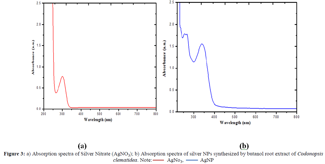
The bio reduction of silver nitrate (AgNO3) to silver NPs was determined periodically by UV-Visible spectroscopy. The sample of synthesized silver NPs was diluted with doubly deionised water and UV-Visible spectrum was recorded by using a quartz cuvette followed by deionized water as a reference. The spectrum was recorded within the range of 200 nm-800 nm.
The UV-Visible spectrum of silver nitrate (AgNO3) shows absorbance at 300 nm at room temperature as shown in Figure 3(a) Whereas the mixture of silver nitrate and butanol root extract of Codnopsisclematidea shows absorption peak around 340 nm as shown in Figure 3(b).
Figure 3. a) Absorption spectra of Silver Nitrate (AgNO3); b) Absorption spectra of silver NPs synthesized by butanol root extract of Codonopsis
clematidea. Note:  AgNO3,
  AgNP
T his change in the absorption spectra of both the solution indicates the formation of silver NPs and similar results were reported [12].
T his change in the absorption spectra of both the solution indicates the formation of silver NPs and similar results were reported [12].
FTIR analysis of silver NPs
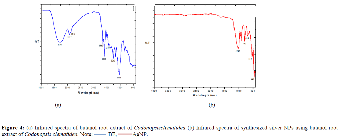
Infrared spectroscopy is used to distinguish between biomolecules for stabilization and capping of the silver NPs which were synthesized using root extract of Codonopsis clematidea [13]. The FT-IR spectrum of the butanol extract of plant is in Figure 4 (a) gives broad band at 3293 cm-1 can be related to the free O-H in a molecule which is not bonded to hydrogen indicates the presence of alcohol and phenol, bands at 2927-2865 cm- 1corresponds to C-H stretch region for the aldehyde, band at 1662 cm-1in the spectra indicates the presence of C=O group, band at 1477-1600 cm- 1characteristic of the C=C stretching bands for aromatic rings, all form plant metabolites [14]. Bands from 1262-1031 cm-1 indicates the primary, secondary or tertiary structure to alcohol respectively. All the above bands indicate the presence of phenolic structure in the butanol extract of plant.
The FTIR spectra of synthesized silver NPs using butanol root extract of Codonopsis clematidea in Figure 4(b) shows, the slight band at 1655 cm-1 which indicates the presence of C=O group, band at 1019 cm-1 shows the C-N stretch, band at the range of 908 cm-1, 793 cm-1, 688 cm-1 gives out of plane bending for =C-H group respectively [14].
Transmission Electron Microscope (TEM) analysis of silver NPs
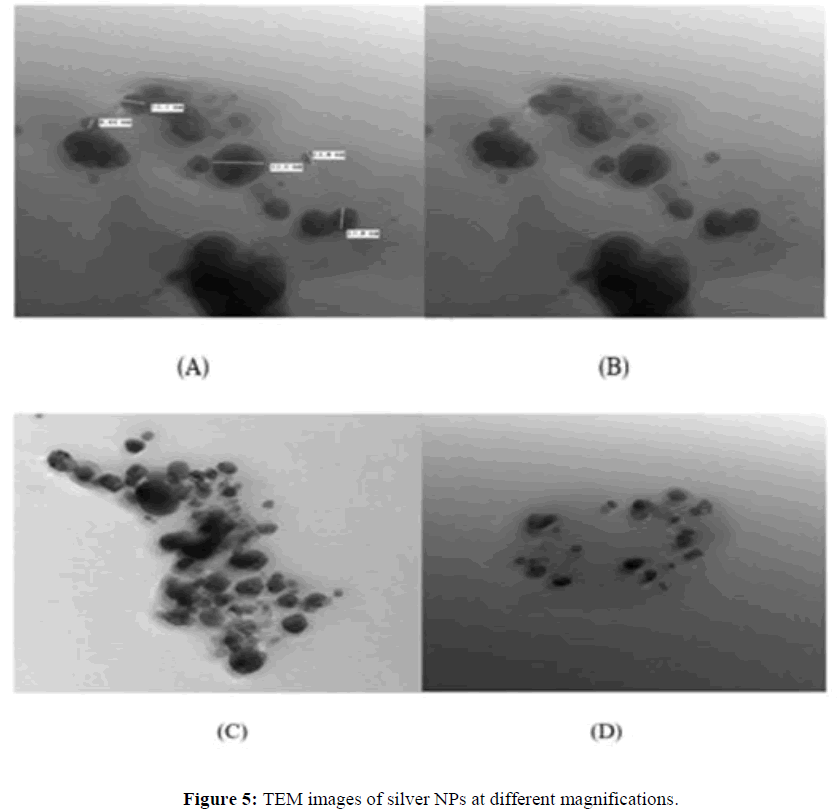
The structural morphology, size and crystallinity of silver NPs were confirmed by TEM analysis micrograph images recorded from different magnifications. TEM micrographs indicate that the synthesized silver NPs were almost spherical and mono-dispersed in nature. Particles were isolated from each other without any agglomeration and having average diameter of ~16 nm. The smallest nanoparticle was of size 8.46 nm and largest was of 32.0 nm as showing in Figure 5 and 6 [15].
X-Ray diffraction (XRD) pattern of silver NPs
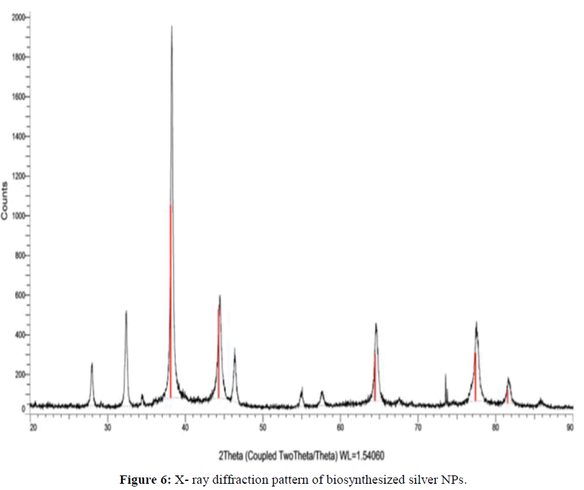
X-ray diffraction (XRD) technique is used for the determination of crystalline characteristics of the synthesized silver NPs. The XRD profile of the bio-synthesized silver NPs by the root extract of Codonopsis clematidea is shown in Figure 5(b). The intense peaks at 38.116, 44.300, 64.445 and 77.399 represents the (111), (200), (220) and (311) Bragg's reflections of the face-centred cubic structure of silver, respectively. However, the diffraction peaks are broad which indicates that small crystalline size is obtained.
Antioxidant potential
The ABTS radical scavenging potential (I%) of different fraction of methanol extract and synthesized silver NPs was compared with vitamin C and gallic acid as a positive control. The butanol fraction had highest scavenging potential as compared to other fraction. The Table 1. Presents I % (ABTS radical scavenging potential) of various fractions and silver NPs. These results may indicates that butanol fraction was rich in polyphenolics pertaining to which it had higher antioxidant potential. This also supported by the fact that only butanol fraction yield fast and good amount of silver NPs as compared to other fractions. Further, the antioxidant potential of silver NPs, butanol and methanol fractions were comparable with the vitamin C and gallic acid used as external standard (Table 1).
| Sr. No. | Name of fraction/NPs | I % (ABTS radical scavenging potential) |
|---|---|---|
| 1 | Hexane fraction | 09.14 |
| 2 | Ethyl acetate fraction | 57.64 |
| 3 | Butanol fraction | 89 |
| 4 | Methanol fraction | 78.34 |
| 5 | Silver NPs | 84 |
| 6 | Vitamin C | 98 |
| 7 | Gallic acid | 96 |
Conclusion
The quick biological synthesis of silver NPs using Codonopsis clematidea root extract offers simple, cost-efficient and eco-friendly greener route for the synthesis of benign metal NPs. The silver NPs are mainly spherical and have average diameter of ~16 nm. The synthesized silver NPs were characterized using UV-Vis, FT-IR, TEM and XRD techniques. Synthesized NPs have comparable antioxidant potential as that of vitamin C and gallic acid (positive control). The literature revealed that the silver NPs synthesized using plant extracts have potential applications in the biomedical area. The present results revealed that the root extracts of Codonopsis clematidea may act as potential source for the green synthesis of various metal NPs. Based on this study, some other NPs may be prepared in future. From the nanotechnology point of view, this is a significant advancement in synthesis of NPs.
References
- Mohammadi S, Pourseyedi S, Amini A. J Environ Chem Eng. 2016, 4: p. 2023-2032.
- Dauthal P, Mukhopadhyay M. Ind Eng Chem Res. 2016.
- Iravani S, Green Chem. 2011, 13: p. 2638-2650.
- Ankamwar DC, Ahmad A, Sastry M. J Nano sci nano technol. 2005, 5: p. 1665-1671.
- Morris KE, Lammers TG. Bot Bull Acad Sin. 1997, 36: p. 277-284.
- Govaerts R. World Checklist of Seed Plants. 1999, 3: p. 1-1532.
- Sharma A, Cannoo DS. RSC Adv. 2016, 6: 78151-78160.
- Sharma A, Cannoo DS. J Food Biochem. 2017, 41: p. 12337-12348.
- Singh J, Dhaliwal AS. Analytical Letters. 2018.
- Raj S, Chand SM, Trivedi R. Biochem Biophy Res Comm. 2018, 503: p. 2814-2819.
- Sadhasivam S, Shanmugan P, Yun K. Colloids Surfaces Bio interfaces. 2010, 81: p. 358-362.
- Dwivedi AD, Gopal K. Colloid Surface Physicochem Eng Aspect. 2010, 369: p. 27-33.
- Kouvaris P, Delimitis A, Zaspalis V, et al. Mater Lett. 2012, 76; p. 18-20.
- Nayak D, Ashe S, Rauta PR, et al. Mater Sci Eng. 2016, 58; p. 44-52.
- Bhardwaj P, Thakur MS, Kapoor S, et al. Pharmacog J. 2019

