Research Article - Der Pharma Chemica ( 2024) Volume 16, Issue 1
Green Synthesis and Evaluation of Pharmaceutical Properties of Euphorbia umbellata (Pax) Bruyns Latex
Padmini AP and Rama Bhat P*Rama Bhat P, Department of PG Biotechnology, Alva's College, Moodbidri, Karnataka, India, Email: bhat_pr@rediffmail.com
Received: 08-Jan-2024, Manuscript No. DPC-24-127546; Editor assigned: 11-Jan-2024, Pre QC No. DPC-24-127546 (PQ); Reviewed: 25-Jan-2024, QC No. DPC-24-127546; Revised: 29-Jan-2024, Manuscript No. DPC-24-127546 (R); Published: 26-Feb-2024, DOI: 10.4172/0975-413X.16.1.232-238
Abstract
Present work explores the synthesis and application of Silver Nanoparticles (AgNPs) using latex from Euphorbia umbellata. The study characterizes the synthesized AgNPs and evaluates their antibacterial, antifungal and larvicidal activities. Phytochemicals in the latex extract are confirmed, including proteins, saponins, phenolics, terpenoids, carbohydrates, alkaloids, steroids and cardiac glycosides. However, flavonoids, tannin and phlobatannins were absent. The characterization of AgNPs involved UV-visible spectroscopy, Fourier Transform Infrared Spectroscopy (FTIR) and Scanning Electron Microscopy (SEM). AgNPs exhibited antibacterial activity against Escherichia coli and Staphylococcus aureus, with increased concentration resulting in larger inhibition zones. They also displayed antifungal activity, particularly against Fusarium solani. In mortality bioassays, AgNPs showed toxicity against Drosophila melanogaster larvae, with 100% mortality in 100 μl/mL concentration within 1 hour. This research highlights the potential of Euphorbia umbellata latex derived AgNPs in various applications, including antimicrobial agents and insect pest control. Further studies may focus on characterizing specific bioactive compounds in the latex.
Keywords
Green synthesis; Silver nanoparticles; Euphorbia umbellata latex; Insect pest control; Antimicrobial activities
Introduction
Nanotechnology, a scientific domain focused on materials at the nanoscale, has led to a convergence with herbal and medicinal plant biology. Technological progress and expanded scientific knowledge have unveiled a unique avenue for research and development. Within this intersection, the green synthesis of plant sourced silver nanoparticles has emerged as a notable interference. Silver nanoparticles possess multifaceted applications across various scientific disciplines, encompassing medicine, pharmacology, electronics, cosmetics, drug delivery systems and cancer therapies. They serve as potent antibacterial agents, found in a wide array of industrial, household and healthcare products. Furthermore, their role in enhancing the efficacy of anti-cancer drugs, particularly in tumor eradication, underscores their significance in nanoparticle based research [1,2]. Euphorbia umbellata (Pax) Bruyns, a native of Africa and a member of the Euphorbiaceae family, is recognized as "Janauba" and "cola-nota" in Brazil. This plant, also called Synadenium grantii, grows up to 3.5 meters in height in its natural habitat. The vast Euphorbia genus encompasses approximately 2000 species, found across temperate and tropical regions, characterized by their latex, known for its poisonous properties [3,4]. The latex is found in specialized cells called “laticifera” throughout the plant. The latex of this species boasts an array of medicinal properties, including antiulcer, anti-inflammatory, homeostatic, antiangiogenic, antiviral activity against hepatitis B, diuretic effects for eliminating kidney stones and prominent antitumor characteristics. In folk medicine, it has been utilized to address a wide spectrum of health issues, such as cancer, allergies, chagas' disease, internal bleeding, sexual impotence, leprosy, obesity, nervous ulcers, menstrual cramps, flu and microbial infections [5,6]. It even showed molluscicidal activity to control schistosomiasis and has been implicated in the defense against herbivorous insects [7]. In the pursuit of sustainable insect pest control for smallholder farmers, plant extracts have emerged as a potential solution. However, their effectiveness in curbing crop damage and impact on plant growth, yield and insect populations remains uncertain. Plant-derived nano pesticides, offering controlled and sustained toxicant release, show promise in effectively reducing pests and plant infestations [8,9,10]. Rising concerns over synthetic pesticides call for eco-friendly, biodegradable alternatives. Plant derived products offer selective and promising solutions in contemporary agrochemical research, safeguarding crops and the environment [11]. The present research aims to evaluate the effect of synthesized nanoparticles on the larvae of Drosophila melanogaster.
Materials and Methods
Sampling of crude latex: Crude latex was collected from Euphorbia umbellata plants from Mysore district, Karnataka, India.
Qualitative phytochemical screening: Aqueous and methanol extracts of latex were screened for various compounds such as proteins, flavonoids, saponins, phenolics, terpenoids, carbohydrates, alkaloids, tannins, steroids, cardiac glycosides and phlobatannins using standard protocols [12].
Green synthesis of silver nanoparticles: Silver nanoparticles were synthesized by reducing silver nitrate using phytochemicals from Euphorbia umbellata latex extract. This involved mixing 3% of 25 mL latex solution with 1 M of 25 mL AgNO3 solution and heating at 80°C for 30 mins to form the nanoparticles [13].
Characterization of synthesized nanoparticles
UV-Visible Spectra analysis: Measuring the absorbance of the synthesized silver nanoparticles.
Fourier Transform Infrared Spectroscopy (FTIR): Identifying functional groups in the nanoparticles.
Scanning Electron Microscope (SEM) analysis: Determining the size and shape of the nanoparticles.
Antibacterial activity: The antibacterial activity of the synthesized silver nanoparticles was tested using the well diffusion assay against various bacteria, including Escherichia coli, Staphylococcus aureus and Pseudomonas aeruginosa using well diffusion assay.
Antifungal activity: The anti-fungal activity was tested using the poison bait method against Fusarium solani and Colletotrichum gloeosporioides [14].
Mortality bioassay: A mortality bioassay was conducted using Drosophila melanogaster larvae, exposed to different concentrations of synthesized nanoparticles and crude latex. The percentage of mortality was calculated based on the number of dead larvae out of the total tested [15].
Results and Discussion
Qualitative phytochemical screening
The aqueous extracts and methanol extracts showed positive results for the majority of the phytochemical tests. The phytochemical tests revealed the presence of proteins, saponins, phenolics, terpenoids, carbohydrates, alkaloids, steroids and cardiac glycosides (Table 1). Screening of E. lateriflora revealed varying important phytochemical components such as alkaloids, saponins, glycoside, steroid, flavonoid, terpenoid, coumarins and reported the absence of tannins in both ethyl acetate and n-hexane [16]. Compounds such as alkaloids, saponins and tannins possess inhibitory potential against bacteria [17]. Saponins have antifungal and molluscicidal activity [18]. The study conducted by Kothale, et al. [19] showed the absence of saponins, tannins and phlobatannins, while flavonols are only present in Jatropha. Presences of alkaloids were common among Euphorbiaceae.
| Phytochemicals | Aqueous extract | Methanol extract |
|---|---|---|
| Proteins | + | - |
| Flavonoids | - | - |
| Saponins | + | + |
| Phenolics | + | - |
| Terpenoids | - | + |
| Carbohydrates | + | - |
| Alkaloids | - | + |
| Tannins | - | - |
| Steroids | + | + |
| Cardiac glycosides | - | + |
| Phlobatannins | - | - |
| Note: +: denotes present, -: denotes absent. | ||
Table 1: Phytochemical screening of aqueous and methanol extract of Euphorbia umbellata latex.
Green synthesis of silver nanoparticles
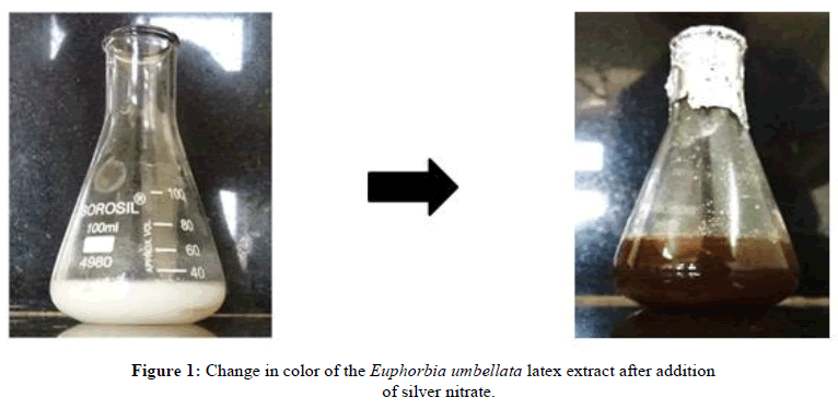
Silver nanoparticles were synthesized by the reduction of silver nitrate by water soluble Euphorbia umbellata latex extract. The latex extract of the plant showed change in color, where the color changed from white to deep brown with the precipitation of the particles during the process of heating at 80°C. The brown color of solution confirmed the reduction of silver nitrates into the silver nanoparticles (Figure 1). The latex of E. nivulia was successfully used to synthesize AgNPs even at high concentrations, the results confirmed the stabilization of the NPs by the latex which gives an advantage for potential biological application and also showed antimicrobial application [20]. The study conducted by Kalaiselvi, et al. [21] showed that 1 and 2 % latex extract are insufficient for nanoparticle synthesis, but increasing the amount of latex extract had a definite influence on the synthesis of AgNPs. They concluded that a 3% latex extract concentration is optimum for the green synthesis of AgNPs.
Characterization of synthesized silver nanoparticles
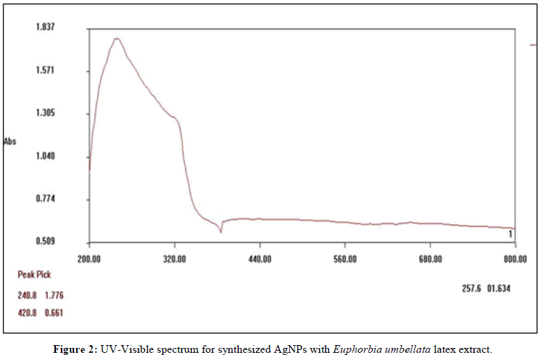
Uv-vis spectral analysis: The synthesis of silver nanoparticles was confirmed by color change followed by UV-visible spectrophotometer analysis. The absorbance spectrum of the colloidal sample was obtained in the range of 200 nm-800 nm, using double beam UV-vis spectrophotometer. The silver nanoparticles are known to exhibit a UV-visible absorption maximum in the range of 400 nm-500 nm due to surface plasmon resonance. The UV-visible spectrophotometer analysis of reaction mixture of silver nanoparticles synthesized using latex extract showed broad peak at 240 nm and sharp peak at 420 nm in the spectrum (Figure 2).
The previous studies reported the characteristic Surface Plasmon Resonance (SPR) absorption band from 424 nm to 437 nm was observed, this increase in indicates increasing size of particles at higher concentrations. Higher concentration of latex or use of external reducing agent did not change the absorbance intensity significantly which ensured complete reduction of Ag+ ions by latex [20]. Another study conducted by utilizing Euphorbia heterophylla leaf extract showed SPR band at 340 nm [22].
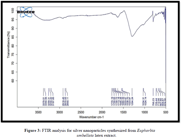
Fourier transform infrared spectroscopy: FTIR analysis of silver nanoparticles was performed in the wave number range of 4000 cm-1-600 cm-1. FTIR spectrum Eu-AgNPs shows absorption peaks at 3379 cm-1 broad peak, O-H (hydroxyl) stretch, indicates alcohol, 2918 cm-1 sharp peak, C-H stretch, indicates alkane functional group, 1770 cm-1 strong peak, C=O stretch, indicates an aldehyde, ketone, ester or carboxylic acid functional group, 1633 cm-1 strong peak, C=C stretch, indicating an alkene functional group (Figure 3). One of the previous studies showed strong broad peak at 3300 cm-1-3500 cm-1 is characteristic of the N-H stretching vibration [23].
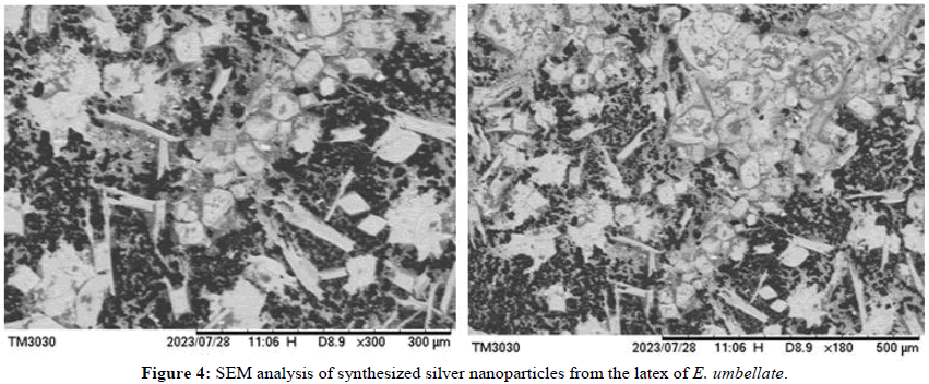
Scanning Electron Microscope (SEM) analysis: The synthesized nanoparticle showed cubic shape and the average size of latex mediated nano particles was found to be 25.84 nm ± 8.9 nm (Figure 4). The studies conducted on Euphorbia tirucalli latex extract showed the nanoparticles of spherical and cubic in the size of 20 nm-30 nm [21].
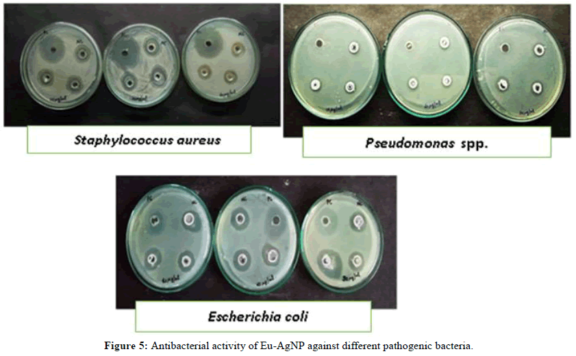
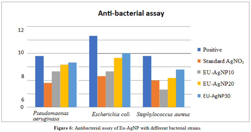
Antibacterial activity by well diffusion assay: Results of the antibacterial activity of the AgNPs showed effects against all three bacterial strains-Escherichia coli, Staphylococcus aureus and Pseudomonas spp. with variation in the diameter of inhibition zones. The maximum zone of inhibition was exhibited against E. coli while the minimum was against Staphylococcus aureus (Table 2 and Figures 5 and 6). Similar results were obtained for different latex extracts of Euphorbia spp. against Staphylococcus aureus, Klebsiella, E. coli and Pseudomonas aeruginosa and the highest inhibition zone was detected against E. coli while the lowest was against Staphylococcus aureus [24].
| Isolate | *Zone of inhibition (mm) | |||
|---|---|---|---|---|
| 10 µl/ml | 20 µl/ml | 30 µl/ml | Negative control | |
| Pseudomonas aeruginosa | 5.3 ± 0.57 | 6.3 ± 0.57 | 6.6 ± 0.57 | 0 |
| Escherichia coli | 5.3 ± 0.57 | 7.3 ±1.15 | 8 ± 1.17 | 0 |
| Staphylococcus aureus | 2.6 ± 0.57 | 4.3 ± 0.57 | 5.6 ± 0.57 | 0 |
| Note: *: mean, ±: standard deviation, N=3. | ||||
Table 2: Antibacterial activity of Eu-AgNP with selected bacterial strains.
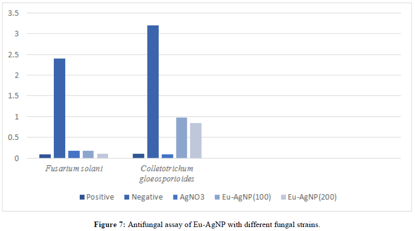
Antifungal activity: The antifungal activity of AgNPs along with the standard AgNO3, bavistin as positive control and distilled water as negative control was tested against Fusarium solani and Colletotrichum gloeosporiodes by poison bait method. The AgNPs had shown better antifungal activity against Fusarium solani when compared to Colletotrichum gloeosporiodes. The Antifungal activity of standard AgNO3 and SNP were compared (Table 3 and Figure 7). Similar studies were carried out by Pruthvi, et al. [25] for the antimicrobial properties of different solvent extracts of E. heterophylla latex. The acetone extract demonstrated a higher zone of inhibition against S. aureus, P. aeruginosa, B. subtilis, A. niger and F. oxyporum.
| Fungal strains | Mycelial dry weight* (g) | ||||
|---|---|---|---|---|---|
| 100 µl/ml | 200 µl/ml | Negative control | AgNO3 | Bavistin | |
| Fusarium solani | 0.18 ± 0.12 | 0.11 ± 0.01 | 1.29 ± 0.005 | 0.06 ± 0.01 | 0.05 ± 0.04 |
| Colletotrichum gloeosporiodes | 0.26 ± 0.04 | 0.14 ± 0.02 | 0.98 ± 0.10 | 0.02 ± 0.05 | 0.02 ± 0.05 |
| Note: *: mean; ±: standard deviation; N=3. | |||||
Table 3: Effect of formulations on the level of lipid peroxidation (MDA), Catalase (CAT) and reduced Glutathione (GSH) in the brain tissue.
Mortality bioassay
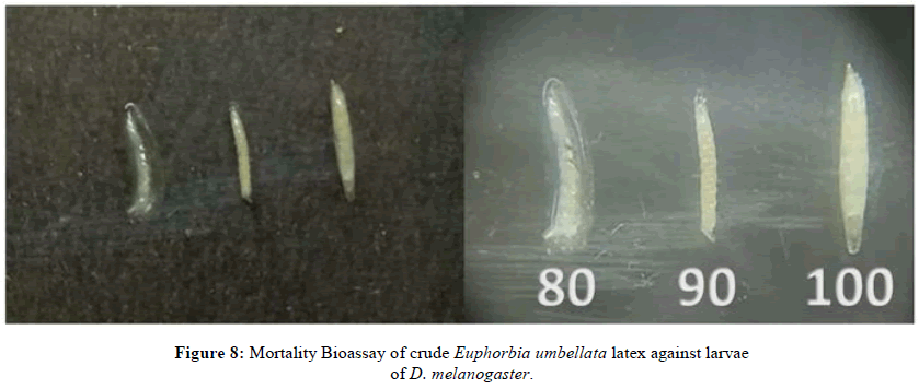
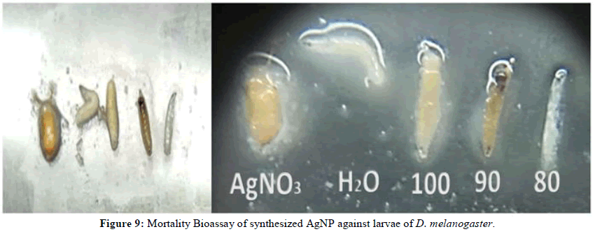
The mortality bioassay data of latex and synthesized AgNP against larvae of Drosophila melanogaster were observed at various concentrations and exposure time. Mortality of Drosophila larvae was observed after time intervals of 1, 5 and 10 h, each concentration was replicated two times. From the observations made, higher concentration of crude latex and synthesized AgNP showed higher mortality in less time. The results of the mortality assay of crude latex and synthesized silver nanoparticles against the larvae are shown in (Tables 4 and 5). It was found that the increased concentrations caused high mortality. The results of lethal concentration of 50% mortality (LC50) using Eu-AgNP and crude latex are shown in (Table 5). E. prostrata and P. hysterophorus showed toxicity against Drosophila melanogaster. Phytochemical constituents showed the presence of flavonoids, saponins, tannins, steroids, cardiac glycosides, alkaloids, anthraquinones and terpenoids (Figures 8 and 9) [15].
| % Mortality | |||||||||
|---|---|---|---|---|---|---|---|---|---|
| Time | 1 hour | 5 hour | 10 hour | ||||||
| Concentrations | 80 µg/ml | 90 µg/ml | 100 µg/ml | 80 µg/ml | 90 µg/ml | 100 µg/ml | 80 µg/ml | 90 µg/ml | 100 µg/ml |
| Feed+water | 0 | 0 | 0 | 0 | 0 | 0 | 0 | 0 | 0 |
| Feed+AgNO3 | 0.0 | 0 | 0 | 0 | 0 | 0 | 0 | 0 | 0 |
| Feed+crude latex | 6.67 | 40 | 73.33 | 13.33 | 60 | 26.66 | 80 | - | - |
| Feed+Eu-AgNP | 6.66 | 66.66 | 100 | 20 | 33.33 | - | 73.33 | - | - |
Table 4: Mortality bioassay of larvae of Drosophila melanogaster.
| LC50 | |||
|---|---|---|---|
| 1 h | 5 h | 10 h | |
| Crude latex | 93 ± 1.6 | 76 ± 1.6 | 65 ± 1.8 |
| Eu-AgNP | 88 ± 1.8 | 35 ± 1.7 | 71 ± 1.8 |
Table 5: Lethal concentration of 50% mortality (LC50) of Eu-AgNP and crude latex.
Conclusion
In conclusion, the synthesis of silver nanoparticles from Euphorbia umbellata latex was successful, as indicated by the color change and confirmed by UV-visible spectroscopy. Characterization using various techniques supported the presence of bioactive phytoconstituents in the latex extract. Phytochemical analysis revealed the existence of proteins, saponins, phenolics, terpenoids, carbohydrates, alkaloids, steroids and cardiac glycosides, while flavonoids, tannins and phlobatannins were absent. FTIR analysis identified functional groups in the synthesized nanoparticles. The silver nanoparticles exhibited antibacterial and antifungal activities, with maximum efficacy against Escherichia coli and Fusarium solani. Additionally, they demonstrated toxicity against Drosophila melanogaster larvae, further highlighting their potential as an insecticide. The presence of diverse phytochemicals underscores the plant's value in traditional medicine and suggests the need for further research to purify and characterize these compounds for potential drug development. Euphorbia umbellata shows promise as an herbal alternative to antimicrobial agents and pest control. Future investigations should focus on harnessing its phenolic compounds for insect pest management programs.
References
- Roy S, Das TK. Int J Plant Biol Res. 2015; 3(3): p. 1044.
- Ebrahimzadeh MA, Tafazoli A, Akhtari J, et al. Anti-Cancer Agents Med Chem. 2018; 18(14): 1962-1969.
[Crossref] [Google Scholar] [PubMed]
- Mohamed NH, Ismail MA, Abdel-Mageed WM, et al. Asian Pac J Trop Biomed. 2014; 4(11): p. 876-883.
- Kumar SA, Priyadarshini HS, Paramita AP, et al. GSC Biol Pharm Sci. 2019; 8(2): p. 35-38.
- Luz LE, Paludo KS, Santos VL, et al. Rev Bras Farmacogn. 2015; 25:p. 344-352.
- Ahmad S, Perveen S, Arshad MA, et al. J Complement Integr Med. 2018; 16(2): p. 201-209.
[Crossref] [Google Scholar] [PubMed]
- Pereira LP, Dias CN, Miranda MV, et al. Rev Inst Med Trop Sao Paulo. 2017; 59(36): p. 85-89.
[Crossref] [Google Scholar] [PubMed]
- Moreau TL, Warman PR, Hoyle J. Biol Agric Hortic. 2006; 23(4): p. 351-370.
- Ndakidemi B, Mtei K, Ndakidemi P. Agric Sci. 2016; 7(6): p. 364
- Gahukar RT, Das RK. Nanotechnol Environ Eng. 2020; 5(1): p. 1-9.
- Hongo H, Karel AK. J Appl Entomol. 1986; 102(15): p. 164-169.
- Shaikh JR, Patil M. Int J Chem Stud. 2020; 8(2): p.603-608.
- Guha P, Mukhopadhyay R, Gupta K. Taiwania Taipei. 2005; 50(4): p. 272.
- Riaz B, Zahoor MK, Zahoor MA, et al. J Evid Based Complementary Altern Med. 2018; 20(12): p.15-18.
[Crossref] [Google Scholar] [PubMed]
- Kamba A, Hassan LG. Afr J Pharm Pharmacol. 2010; 4(9): p. 645-652.
- Doughari JH, Okafor NB. Afr J Microbiol Res. 2008; 2(1): p. 042-046.
- Aboaba OO, Efuwape BM. Bio Res Comm. 2001; 13:p. 183-188.
- Kothale KV, Rothe SP, Pawade PN. J Phytol. 2011; 3(12): p. 60-62.
- Valodkar M, Nagar PS, Jadeja RN, et al. Colloids Surf A: Physicochem Eng. 2011; 384(1-3): p. 337-344.
- Kalaiselvi D, Mohankumar A, Shanmugam G, et al. J Crop Prot. 2019; 117: p. 108-114.
- Rosa RL, Campos PM, Cruz LS, et al. Res Soc Dev. 2022; 11(12): p. 1123-1125.
- Jin L, Bai R. Langmuir. 2002; 18(25): p. 9765-9770.
- Saleh GM, Najim SS. Iraqi J Sci. 2020: 14(7): 1579-1588.
- Ml PR, Mk MA, Mary RS. Asian J Pharm Clin Res. 2020; 13 (7): p. 141-145.
- Khan S, Taning CN, Bonneure E, et al. Phytoparasitica. 2017; 45: p. 113-124.

