Research Article - Der Pharma Chemica ( 2021) Volume 13, Issue 10
GC.MS Analysis and Immuo Modulatory Activities of Punica granatum (Cortex) and Zingiber officinales (rhizome)
Esraa M.M. Ali1,2* and Hasan M.H. Muhaisen12Animal Resources Research Corporation - Ministry of Livestock - Fisheries and Rangelands, Sudan
Dr. Esraa M.M. Ali, Department of Chemistry, Faculty of Science and Arts, Najran University, Sharurah, Kingdom of Saudi Arabia, Email: esraamusa28@gmail.com
Abstract
This study was designed to investigate the pharmacotoxicity of methanolic extracts of Punica granatum cortex and Zingiber officinales rhizome at different doses 800,400 and 200mg/kg to rats via oral route, The methanolic extract of Punica granatum cortex (400 mg/kg+ PHA) in days 45 showed significant increase in IL-2 and IL-6 level. While the methanolic extract of Zingiber officinales rhizome (400 mg/kg+ PHA) in days 45, showed significant increase in IL-10 level.
Keywords
Zingiberaceae, lythraceae, Cytokine, Interleukins and Interferon INFγ.
Introduction
Medicinal plants are most important to daily health and practices particularly in developing countries, also became a topic of augmented global importance, having impacted on both world health and international trade.
Man has always made use of flora to alleviate suffering. It is well known that many medicinal plants are useful in a crude form, for example: Atropa belladona as an antiseptic, Rauvolia serpentine root as an anti-hypertension and as tranquilizer, Papever somniferum extract or tincture as analgesic (Oliver- Bever, 1986).
Pomegranate is used in several systems of medicine for a variety of ailments. In Ayurvedic medicine the pomegranate is considered “apharmacy unto itself” and is used as an antiparasitic agent, [1] as “blood tonic” [2] and to heal aphthae, diarrhea, and ulcers [3].
The chemical constituents were separated by silica gel column chromatography and their structures were determined by spectroscopic experiments. As a result, twelve compounds were isolated and identified as oleanolic acid, ursolic acid, palmitic acid, tricin, catechin, rutin, apigenin, apigenin-7-O-glucoside, 2S, 3S, 4S-trihydroxypentanoic acid, gallic acid, β-sitosterol, and daucosterol [4].
It was indicated that alkaloid was present at the rate of 0.35 to 0.60% in the body rinds, and over 3% in the roots; but none was found in the fruit rinds [5]. It was also indicated that pseudopelletierine, pelletierine, isopelletierine, methylpelletierine 1-pelletierine, dl-pelletierine and methyl isopelletierines were found in composition of the root, body and branch rinds of P. granatum [6,7]. It was detected that saturated alkaloids present in the root and body rinds are not present in the leaves, whereas (2-2-propenyl)-piperidine of unsaturated alkaloids was present in the leaf extract [8]. Tannin and similar compounds were stated that Punica cortein A, B, C, D in the structure of hydrolysable C-glycoside, which is a new ellagitannin, as well as punigluconin which contains one gluconic acid; and also, casuariline and casuarine were present in the fresh body roots of P. granatum [9].
Medicinal uses of ginger has been claimed to decrease the pain from arthritis, though studies have been inconsistent. It may also have blood thinning and cholesterol lowering properties that may make it useful for treating heart disease. Preliminary research also indicates that nine compounds found in ginger may bind to human serotonin receptors, possibly helping to affect anxiety [10].
The characteristic odor and flavor of ginger is caused by a mixture of Zingerone, Shogaols and Gingerols, volatile oils that compose one to three percent of the weight of fresh ginger. In laboratory animals, the [6]-gingerols increase the motility of the gastrointestinal tract and have analgesic, sedative, antipyretic and antibacterial properties [11]. Ginger oil has been shown to prevent skin cancer in mice and a study at the University of Michigan demonstrated that gingerols can kill ovarian cancer cells [13-14]. [6]-Gingerol [1-4'-hydroxy-3'-methoxyphenyl]-5-hydroxy-3-decanone) is the major pungent principle of ginger. The chemopreventive potentials of [6]-gingerol present a promising future alternative to expensive and toxic therapeutic agents [15].
Cytokine super families contain important regulatory cell membrane receptor-ligand pairs, reflecting evolutionary pressures that use common structural motifs in diverse immune functions in higher mammals. The TNF/TNF receptor super family 1contains cytokines, including TNF- α,; lymphotoxins and cellular ligands, CD40L, mediates B cell and T cell activation, and FasL (CD95),which promotes apoptosis). Similarly, the IL-1/IL-1 receptor super family- 2 contains cytokines, including IL-1 -β, IL-1 -α, IL-receptor antagonist, IL-18, and IL-33, which mediate physiologic and host-defense function, but this family also includes the Toll-like receptors, a series of mammalian pattern-recognition molecules with a crucial role in recognition of microbial species early in innate responses [16]. Interleukin -2 (IL-2) proliferation is essentially a regulator as it is product of T cells which acts primary on T cells. Interleukin -4 (IL-4) also a T cells product, appears to be multi –specific, acts on T cells, B cells and macrophages and is probably essential for the maintenance of an activated immune response in vivo. Interleukin-6 (IL-6) has been relatively recently assigned; human IL-6 was the first to be identified and is known by other names including interferon β2 (IFN-β2) and B lymphocyte –stimulating factor- 2 (BSF-2). They are three types of interferon: interferon α, β and γ. Interferon α, (leucocyte) interferon, interferon β, (fibroblast) interferon and interferon γ, (immune interferon), were first recognized for their ability to confer resistance to viral infection. Their major functions are; anti-viral, anti-tumor, regulate differentiation, modulate lipid metabolism, inhibits angiogenesis, immunoregulations, (monocyte/macrophage activation) and enhances MHC expression class I: IFN- α and IFN- β, class II: IFN- γ.
Materials and Methods
Plant material
Punica granatum cortex and Zingiber officinales rhizome were purchased from Khartoum North market (Attarin), Khartoum, Sudan. They were identified by the (Dr. Eltayeb Fadul) and the voucher specimen (654) was deposited in the herbarium of (Medicinal and Aromatic plant Institute).
Extraction of Plant Material
The plant materials were dried and powdered. The powdered samples were extracted by methanol using soxhellet apparatus and the obtained Punica granatum methanol extract (PGME) as well as Zingiber officinales methanol extract (ZOME) were concentrated to dryness using rotatory evaporator under reduced pressure and low temperature (<40Cº). The obtained dried extract was kept in airtight containers and stored at low temperature for further studies.
Phytochemical Studies
Gas Chromatography-Mass Spectrometry (GC-MS) analysis of PGME and ZOME
Gas Chromatography, Mass Spectrometer (GC.MS): Chemical composition of PGME and ZOME were determined by using GC-MS (Shomadzu, Japan, TQ8040), fitted with Flame ionization detector (FID). Separation of sample was achieved using C18 column, (serial number: us6551263H) having 0.25um film thickness, 30 meter length, and 0.25 mm inner diameter. The injection temperature was set at 250.00ºC and 122.0 KP a pressure. The carrier gas used was hydrogen and given in split mode. Twenty milligrams each of the crude extracts were dissolved in 5 ml of HPLC grade methanol and filtered through 0.22 μm membrane filter. One microliter of the test extract was injected into the system using specific syringe. The identity of the compounds was confirmed by comparing mass spectral data from National Institute of Standards and Technical 2008 library.
Immuno-modulation Studies: The effect of PGME and ZOME was assessed on serum cytokines of albino rats. The experimental protocol was approved by the Institutional Animal Ethics Committee as per the approved Guidelines of Committee for the Purpose of Control and Supervision of Experiments on Animals. Experimental rats were maintained in the Departmental Animal House and provided ad libitum pelleted feed. Rats had free access to clean drinking water and light and dark cycle of almost 12 h was maintained.
The panel of validated assays and experimental approaches used in almost all modern immunopharmacology assessment are resulted from the seminal works of [17] as follows
a) Pyto Heamato Agglutinin (PHA) Protocol
b) Monitoring the Cytokines level with ELISA
Experimental animals
A total of 100 Wister albino male and female rats used were housed in hygienic fiber glass cages. Animals were maintained in 10 group of 10 rat each. Rats were apparently healthy and weighed 90-120 g. Rats were fed on commercial pellets. The rats were allowed free access to drinking water and feed throughout the experimental study. All animals were observed daily for any clinical physiology behavior changes. 8 out of 10 groups were treated with PGME and ZOME respectively. The remaining groups were considered as control by drinking tap water only. The total period of the experimental continued for 45 days. At each time point, 5, 30 and 45 days.
Pyto Heamato Agglutinin (PHA) Protocol
0.5 mg powder of PHA dissolved in 100 ml distill water according to the protocol. Groups which received PHA taken single dose S/C at 0.005mg/kg.
• Group (1): was used as control negative untreated.
• Group (2): was used as positive group stimulant with 0.5 ml Phyto Heamato Aglotinin (PHA).
• Group (3): was treated with PGME (400mg/kg/bw/day) for 45 days and stimulant with PHA
• Group (4): treated with PGME (200mg/kg/bw/day) for 45 days and stimulant with PHA
• Group (5): was treated with PGME (400mg/kg/bw/day) for 45 days.
• Group (6): was treated with PGME (200mg/kg/bw/day) for 45 days.
• Group (7): was treated with ZOME (400mg/kg/bw/day) for 45 days and stimulant with PHA.
• Group (8): was treated with ZOME (200mg/kg/bw/day) for 45 days and stimulant with PHA.
• Group (9): was treated with ZOME (400mg/kg/bw/day) only for 45days.
• Group (10): was treated with ZOME (200mg/kg/bw/day) only for 45 days.
Monitoring the Cytokines level with ELISA
The cytokines, IL-2, IL-4, IL-6, IL-10, IFN-y levels were monitored using Quantkine mouse cytokines immunoassay kit (P&D system, USA). According to manufacturer direction [18].
• Prepare all the reagent and samples as directed in the previous sections.
• Determine the number of wells to be used and put any remaining wells and the desiccant bank into the pouch and seal the Ziploc, store unused well at 4Co.
• Set bank well without any solution.
• Add 100 μL of standard or samples per well. Standard need test in duplicate.
• Add 100 μL of HRP- conjugate to each well (not to blank well). Mix well and then incubate for 2 hours at 37Cº.
• Aspirate each well and wash, repeating the process tow times for a total of three washes. Wash by filling each well with Wash Buffer (200 μ) using a quirt bottle, multi- channel pipette, manifold dispenser, or auto washer, and let it stand for 10 seconds, complete removal of liquid at each step is essential to good performance. after the last wash, remove any remaining wash buffer by aspirating or decanting. Invert the plate and bolt it against clean paper towels.
• Add 50 μL of substrate A and 50 μl substrate B to each well, mix well. Incubate for 15 min at 37Cº. keeping the plate away from drafts and other temperature fluctuations in the dark.
• Add 50 μL of stop solution to each well, gently tap the plate to ensure thorough mixing.
• Determine the optical density of each well within 10 minutes, using a microplate reader set to 450 nm.
Briefly,50 μg/ml of the assay of IL-2, IL-4, IL-6, IL-10, IFN-y levels diluents, were added to each well. Then 50μg of standard control and samples were added. After 2h incubation a 100 μg of the conjugate were added to each well after thorough washing and incubated for overnight. After thorough washing, a 100 μL of the substrate solution was added to each well and incubated for 30 min. Finally, a 100 μg of the solution was added to each well and optical density was measured using ELISA reader with dual wavelength 450 and 750 nm (Thermo Labsystem, Finland).
Statistical analysis
The significance of differences between means was compared at each time point using Duncan’s multiple range test after ANOVA for one-way classified data [19,20].
All data were presented as means (± S.D). Statistical analysis for all. The assays results were done using Microsoft excel program. Student T. test was used to determined significant difference between control and plant extracts at level of (P < 0.5, P < 0.05 and P < 0.01).
The Student-test and ANOVA, one-factor, were used for the analysis of the cytokines level at different immune cells sub-population and the level of cytokines. The Duncan multiple range test (DMRT), which is a reasonable multiple comparison test was used to analyze the differences between the levels of cytokines at different time intervals. The test is designed to compare population means by multiple comparisons of sample means. The data will be analyzed with two-factor analysis of variance followed by Duncan multiple range test.
Results
Phytochemical studies
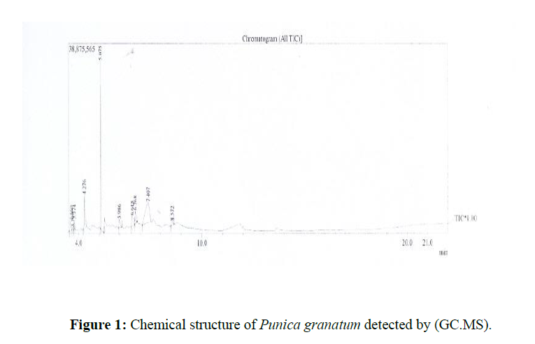
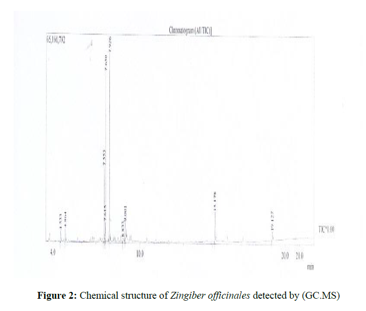
Chemical structure of compounds presents in PGME and ZOME were detected by Gas Chromatography Mass Spectrometer (GC.MS). The chemical constituents detected in PGME were:5hydroxymethylfuraldehyde, laxiric acid, mannitol-manna sugar, oleanolic acid, palmitic acid, gallic acid and ellagic acid ( Table (1); Figure (1) while the chemical constituents found in ZOME were zingiber, alpha-aromadendrene, borneol, β-sesquiphellandrene, gingerols and shogaol Table (2); Figure (2).
| Name | Structure | Formula | Molecular weight |
|---|---|---|---|
| Punica granatum. | |||
| 5-Hydroxymethylfuraldehyde |  |
C6H6O3 | 126.11004 g/mol |
| Laxiric Acid | 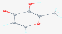 |
C6H6O3 | 126.11004 g/mol |
| Mannitol-manna sugar |  |
C6H14O6 | 182.17176 g/mol |
| Oleanolic acid | 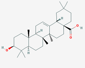 |
C30H48O3 | 456.71 g/mol |
| Palmitic acid |  |
C16H32O2 | 256.4241 g/mol |
| Gallic acid | 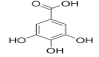 |
C6H6OH3 | 170.11954 g/mol |
| Ellagic acid |  |
C14H6O8 | 302.197 g/mol |
| Name | Structure | Formula | Molecular weight |
|---|---|---|---|
| s rhizome | |||
| Zingiberene |  |
C15H24 | 204.35106 g/mol |
| Alpha-Aromadendrene | 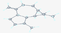 |
C15H24 | 204.35106 g/mol |
| Borneol |  |
C10H18O | 154.24932 g/mol |
| ß-Sesquiphellandrene |  |
C15H24 | 204.35106 g/mol |
| Gingerols | 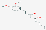 |
C17H26O4 | g/mol 294.38 |
| Shogaol |  |
C17H24O3 | g/mol 276.38 |
The effect of PGME and ZOME on some serum cytokines
Rats were administered PGME and ZOME daily with doses of 400 and 200 mg/kg and their influence were analyzed on the cytokines, IL-2, IL-4, IL-10, and IFN-γ.
The levels of cytokines concentration measured by ELISA test are summarized in tables. The two-factor analysis of variance of cytokines that were monitored in cells of treated rats were either pulsed with Phyto Heamato Aglotinin (PHA) or grown in medium free of PHA indicated a significant difference with time and response. Rat’s cells pulsed with PHA and non-pulsed cells expressed a significant increase in their levels as measured by ELISA on days 5, 30, and 45.
The cytokines model treated with PGME
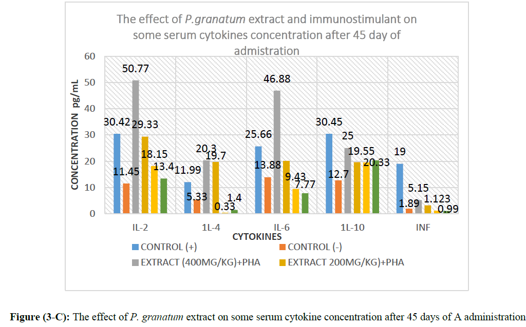
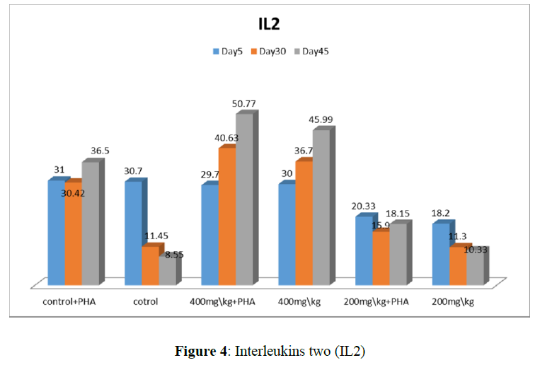
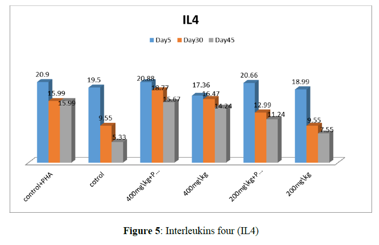
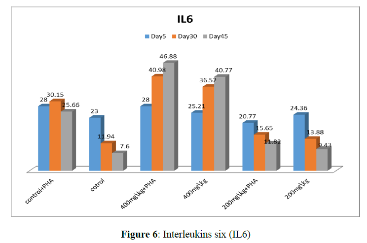
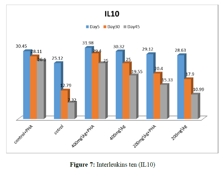
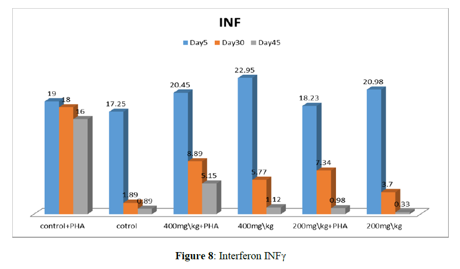
The PGME (400 mg/kg+PHA) showed significant increase in IL-2 (50.77) and IL-6 (46.88) level in day 45 summarized tables (3-AC) and Figure (3-ABC, 4, 5, 6, 7, and 8).
| Days 5 | IL2 (pg/ml) | IL4 (pg/ml) | IL6 (pg/ml) | IL10 (pg/ml) | INF γ (pg/ml) |
|---|---|---|---|---|---|
| Control+ PHA | 31.99 ± 0.44* | 20.49 ± 0.1NS | 26.54 ± 0.4NS | 30.45 ± 0.78* | 19.00 ± 0.00NS |
| Control | 28.66 ± 0.89 NS | 19.99 ± 0.65 NS | 25.00 ± 0.25 NS | 25.96 ± 0.1 NS | 17.25 ± 0.69 NS |
| Extract(400mg\kg)+PHA | 27.32 ± 0.56 NS | 20.88 ± 0.98 NS | 20.24 ± 0.36 NS | 29.6 ± 60.52 * | 22.63 ± 0.58 NS |
| Extract(400mg\kg) | 27.66 ± 0.21 * | 17.66 ± 0.78 NS | 25.00 ± 0.96 NS | 30.04 ± 0.03 * | 19.85 ± 0.47 NS |
| Extract 200mg\kg)+PHA | 30.55 ± 0.45 NS | 19.37 ± 0.45 NS | 15.77 ± 0.85 NS | 29.12 ± 0.7 NS | 18.23 ± 0.47 * |
| Extract (200mg\kg) | 25.1 ± 0.74 NS | 19.37 ± 0.12 NS | 20.99 ± 0.74 NS | 17.09 ± 0.00 | 20.04 ± 0.00 NS |
Key
G1=Control+PHA.
G2=Control.
G3= 400mg/kg Punica granatum+ PHA.
G4=400mg/kg Punica granatum.
G5=200 mg/kg Punica granatum+ PHA. G4=200mg/kg Punica granatum.
Values are mean ± SE. NS=not significant, *P< 0.05, **P<0.01, ***P<0.001.
| Days 30 | IL2 (pg/ml) | IL4 (pg/ml) | IL6 (pg/ml) | IL10 (pg/ml) | INF γ (pg/ml) |
|---|---|---|---|---|---|
| Control+ PHA | 26.44 ± 0.12** | 11111115.99 ± 0.12* | 30.15 ± 0.11* | 30.15 ± 0.00* | 18.00 ± 0.78* |
| Control | 11.45 ± 0.32 NS | 5.33 ± 0.45 NS | 15.6 ± 0.5 NS | 12.79 ± 0.00 NS | 0.89 ± 0.7 NS |
| Extract(400mg\kg)+PHA | 30.1 ± 0.01** | 23.15 ± 0.63* | 40.98 ± 0.1* | 30.74 ± 0.5*** | 8.89 ± 0.32* |
| Extract(400mg\kg) | 22.4 ± 0.5** | 16.47 ± 0.1* | 20.05 ± 0.2* | 33.19 ± 0.1*** | 5.77 ± 0.12 NS |
| Extract 200mg\kg)+PHA | 20.33 ± 0.4** | 1.6 ± 0.09* | 11.66 ± 0.77 NS | 27.25 ± 0.02** | 1.89 ± 0.41 NS |
| Extract (200mg\kg) | 15.00 ± 0.05 NS | 3.09 ± 0.7 NS | 5.77 ± 0.52 NS | 17.23 ± 0.14 NS | 3.05 ± 0.00 NS |
Key
G1=Control+PHA.
G2=Control
G3= 400mg/kg Punica granatum+ PHA
G4=400mg/kg Punica granatum.
G5=. 200mg/kg Punica granatum+ PHAG4=200mg/kg Punica granatum.
Values are mean ± SE. NS=not significant, *P< 0.05, **P<0.01, ***P<0.00
| Days 45 | IL2 (pg/ml) | IL4 (pg/ml) | IL6 (pg/ml) | IL10 (pg/ml) | INFγ (pg/ml) |
|---|---|---|---|---|---|
| Control+ PHA | 30.560.78* | 1199 ± 0.32NS | 25.66 ± 0.11NS | 30.45 ± 0.44* | |
| 19.00 ± 0.01NS | |||||
| Control | 11.45 ± 0.02 NS | 5.33 ± 0.82 * | 13.88 ± 0.19 * | 12.07 ± 0.01 NS | 1.89 ± 0.84* |
| Extract(400mg\kg)+PHA | 50.77 ± 0.13* | 20.03 ± 0.71 NS | 46.88 ± 0.26 NS | 25.00 ± 0.7** | 5.15 ± 0.51 NS |
| Extract(400mg\kg) | 29.33 ± 0.46* | 19.07 ± 0.93 NS | 20.13 ± 0.48 NS | 19.55 ± 0.42** | 1.12 ± 0.62 NS |
| Extract 200mg\kg)+PHA | 18.15 ± 0.79* | 0.33 ± 0.05 NS | 9.43 ± 0.35 NS | 19.55 ± 0.53** | 0.99 ± 0.87* |
| Extract (200mg\kg) | 13.04 ± 0.31 NS | 1. 4 ± 0.44 * | 7.77 ± 0.24 * | 20.33 ± 0.01 NS | 0.99 ± 0.45* |
Key
G1=Control+PHA
G2=Control.
G3= 400mg/kg Punica granatum+ PHA
G4=400mg/kg Punica granatum.
G5=. 200mg/kg Punica granatum+ PHA.G4=200mg/kg Punica granatum.
Values are mean ± SE NS=not significant, *P< 0.05, **P<0.01, ***P<0.001.
Result of 5 days
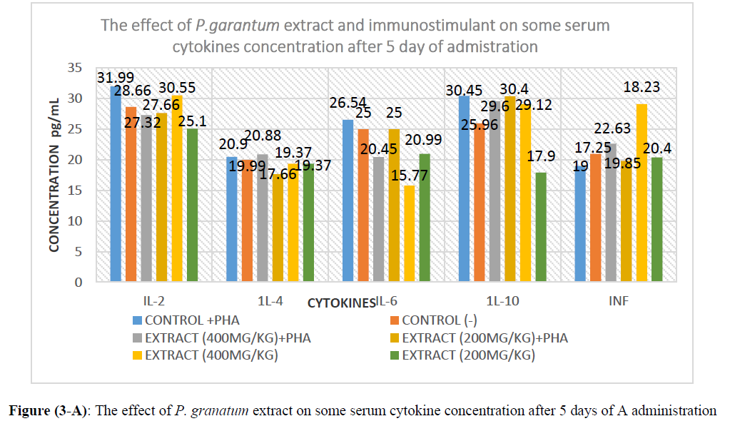
IL-2: the result of group 1 which received PHA at 0.5 I/M showed 31.99 pg/ml, while the control Normal saline, group (2) recorded 28.66 pg/ml. Group 3 which received PGME (400 mg) with PHA showed 27.32 pg/ml. The group 4 which received the PGME (400 mg) only recorded 27.66 pg/. However group 5 received PGME (200 mg) with PHA recorded 30.55 pg/ml and group 6 which received PGME (200 mg) only recorded 25.1 pg/ml IL-2, 19.37 pg/ml IL-4, 20.99 pg/ml IL-6, 17.9 pg/ml IL-10 and 20.4 pg/ml INFγ. (Table 3-A; Figure 3-A).
IL-4: group (1) recorded 20.9 which group (2) recorded 19.99.And group (3) 20.88. However, group 4, 5 and 6 recorded 17.66, 19.37 and 19.37, respectively.
IL-6: Group 1,2,3,4,5 and 6 recorded 26.54, 25, 20.45, 25, 15.77 and 20.99 respectively.
IL-10: Group 1,2,3,4,5 and 6 recorded 30.45, 25.96, 29,6, 30.4, 29.12 and 17.9 respectively.
INFγ: Group 1,2,3,4,5 and 6 recorded 19.00, 17.25, 22.63, 19.85, 18.23 and 20.4 respectively.
Result of day 30
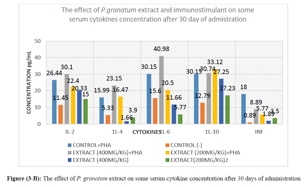
IL-2: the group 1 which received PHA at 0.5 I/M showed 26.44 pg/ml. While the control Normal saline, group (2) recorded 11.45 pg./ml. However Group 3 which received PGME (400 mg) with PHA showed 30.1 pg/ml. Group 4 which received the PGME (400 mg) only recorded 22.4 pg/ml. However, group 5 received PGME (200 mg) with PHA recorded 20.33 pg/ml and group 6 which received PGME (200 mg) only recorded 15.00 pg/ml. Showed in table no (3-B) and figure no (3-B).
IL-4: Group 1,2,3,4,5 and 6 recorded 15.99, 5.33, 23.15, 16.47, 1.6 and 3.9 respectively.
IL-6: Group 1,2,3,4,5 and 6 recorded 30.15, 15.6, 40.98, 20.5, 11.66 and 5.77 respectively.
IL-10: Group 1,2,3,4,5 and 6 recorded 30.15, 12.79, 30.74, 33.12, 27.25, and 17.23 respectively.
INFγ: Group 1,2,3,4,5 and 6 recorded 18.00, 0.89, 8.89, 5.77, 1.89 and 3.5 respectively.
Result of day 45
IL-2: the group 1 which received PHA at 0.5 I/M showed 30.42 pg/ml. While the control Normal saline, group (2) recorded 11.42 pg./ml. However Group 3 which received PGME (400 mg) with PHA showed 50.77 pg/ml. Group 4 which received the PGME (400 mg) only recorded 29.33 pg/ml. However, group 5 received PGME (200 mg) with PHA recorded 18.15 pg/ml and group 6 which received PGME (200 mg) only recorded 13.04 pg/ml. showed in table no (3-C) and figure no (3-C).
IL-4: Group 1,2,3,4,5 and 6 recorded 11.99, 5.33, 20.3, 19.7, 0.33 and 1.4 respectively.
IL-6: Group 1,2,3,4,5 and 6 recorded 25.66, 13.8, 46.8, 20.13, 9.43 and 7.77 respectively.
IL-10: Group 1,2,3,4,5 and 6 recorded 30.45, 12.7, 25, 19.55, 19.55 and 20.33 respectively.
INFγ: Group 1,2,3,4,5 and 6 recorded 19.00, 1.89, 5.15, 1.12, 0.99 and 0.99 respectively.
The cytokines model treated with ZOME
Cytokine levels in the methanolic extract of Zingiber officinales rhizome, treated with 400 and 200mg/kg/bw in days 5 significant (P ≥0.0002) increase in the level of cytokines in all groups
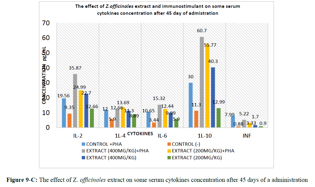
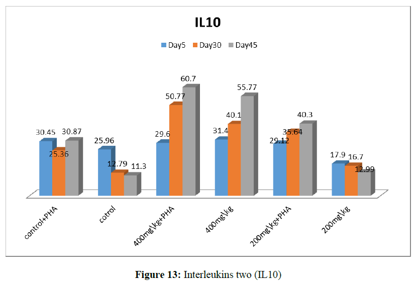
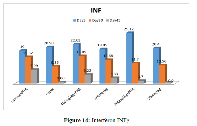
Days 30 and 45 showed significant (P≥0.0002) increase the level of cytokines IL-10 (50.7-60.7) in groups 4 extract +PHA, rats received with doses 400mg/kg/bw/day daily for 45 days were stimulant with PHA. Summarized in table (4-ABC) and figure no (9-ABC.10, 11, 12, 13 and 14).
| Day 5 | IL2 (pg/ml) | IL4 (pg/ml) | IL6 (pg/ml) | IL10 (pg/ml) | INF γ (pg/ml) |
|---|---|---|---|---|---|
| Control +PHA | 31.99 ± 0.065NS | 20. 9 ± 33 NS | 26.54 ± 0.74NS | 30.45 ± 0.06 NS | 19.00 ± 0.03* |
| Control | 28.66 ± 0.21 NS | 19.99 ± 0.1* | 25 ± 0.03* | 25.96 ± 0.06 NS | 20.98 ± 0.14* |
| Extract(400mg\kg)+PHA | 27.32 ± 0.9 NS | 20.88 ± 0.4* | 16.32 ± 0.87 NS | 29.6 ± 0.78 NS | 22.63 ± 0.89 |
| Extract(400mg\kg) | 31.55 ± 0.87 NS | 15. 9 ± 0.7* | 25.12 ± 0.69 NS | 30.40 ± 0.14 NS | 24.32 ± 0.78* |
| Extract(200mg\kg)+PHA | 27.66 ± 0.24 * | 17.99 ± 0.75* | 15.77 ± 0.79* | 29.12 ± 0.36 NS | 19.8 ± 0.45* |
| Extract (200mg\kg) | 25.1 ± 0.96* | 17.99 ± 0.72* | 18.92 ± 0.02* | 17.9 ± 0.4 NS | 25.36 ± 0.12* |
Key
G1=Control+PHA.
G2=Control.
G3= 400mg/kg Zingiber officinale+ PHA
G4=400mg/kg Zingiber officinale
G5=. 200mg/kg Zingiber officinale + PHA.G4=200mg/kg Zingiber officinale.
Values are mean ± SE. NS=not significant, *P< 0.05, **P<0.01, ***P<0.001.
| Day 30 | IL2 (pg/ml) | IL4 (pg/ml) | IL6 (pg/ml) | IL10 (pg/ml) | INF γ (pg/ml) |
|---|---|---|---|---|---|
| Control +PHA | 26.44 ± 0.12** | 15.99 ± 0.87* | 19.87 ± 0.47** | 25.36 ± 0.58** | 15.32 ± 0.14* |
| Control | 11.45 ± 0.79 NS | 10.36 ± 0.17 NS | 10. 07 ± 0.14 NS | 12.79 ± 0.45* | 9.86 ± 0.24 NS |
| Extract(400mg\kg)+PHA | 30.10 ± 0.45 ** | 18.09 ± 0.01* | 17.11 ± 0.09** | 50.77 ± 0.69 ** | 15.89 ± 0.85 NS |
| Extract(400mg\kg) | 22.40 ± .05** | 16.47 ± 0.34* | 12.77 ± 0.14** | 40.01 ± 0.36** | 13.68 ± 0.71 NS |
| Extract 200mg\kg)+PHA | 20.33 ± 0.71* | 15.77 ± 0.07* | 11.79 ± 0.89* | 35. 46 ± 0.74* | 11.07 ± 0.52 NS |
| Extract (200mg\kg) | 15.00 ± 0.47 NS | 12.11 ± 0.97 NS | 13.88 ± 0.31 NS | 16.07 ± 0.85* | 10.56 ± 0.63 NS |
Key
G1=Control+PHA.
G2=Control
G3= 400mg/kg Zingiber officinale+ PHA
G4=400mg/kg Zingiber officinale.
G5=. 200mg/kg Zingiber officinale + PHA.G4=200mg/kg Zingiber officinale
Values are mean ± SE. NS=not significant, *P< 0.05, **P<0.01, ***P<0.001.
| Day 45 | IL2 (pg/ml) | IL4 (pg/ml) | IL6 (pg/ml) | IL10 (pg/ml) | INF γ (pg/ml) |
|---|---|---|---|---|---|
| Control+ PHA | 19.56 ± 0.52** | 12.00 ± 0.12* | 10.65 ± 0.32* | 30.00 ± 0.78* | 7.99 ± 0.32* |
| Control | 9.35 ± 0.36 NS | 5.09 ± 0.34 NS | 3.44 ± 0.45 NS | 11.13 ± 0.98 NS | 0.88 ± 0.49 NS |
| Extract(400mg\kg)+PHA | 35.87 ± 0.98** | 12.66 ± 0.12* | 12.32 ± 0.79** | 60.07 ± 0.1* | 5.22 ± 0.3 NS |
| Extract(400mg\kg) | 24.99 ± 0.32** | 13. 69 ± 0.12* | 12.44 ± 0.4** | 55.77 ± 0.44* | 3.33 ± 0.14 NS |
| Extract 200mg\kg)+PHA | 22.07 ± 0.07* | 11.03 ± 0.09 NS | 9.99 ± 0.87 NS | 40.30 ± 0.77* | 1.07 ± 0.78 NS |
| Extract (200mg\kg) | 12.66 ± 0.7 NS | 8.89 ± 0.87 NS | 5.09 ± 0.65 NS | 12.99 ± 0.64 NS | 0.9 ± 0.00 NS |
Key:
G1=Control+PHA.
G2=Control.
G3= 400mg/kg Zingiber officinale+ PHA.
G4=400mg/kg Zingiber officinale.
G5= 200mg/kg Zingiber officinale + PHA.G4=200mg/kg Zingiber officinale.
Values are mean ± SE. NS=not significant, *P< 0.05, **P<0.01, ***P<0.001.
Result of 5 days
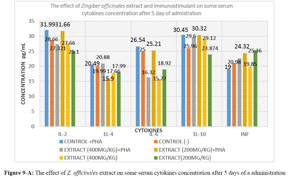
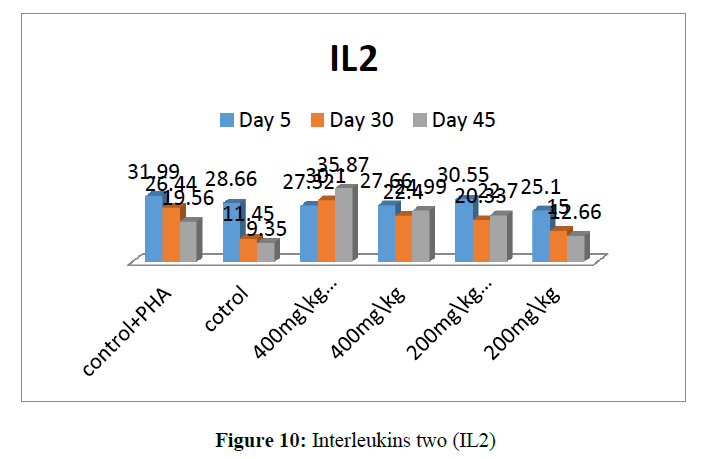
IL-2: the result of group (1) which received PHA at 0.5 I/M showed 31.99, while the control normal saline. Group (2) recorded 28.66. The group (3) which received 400mg of the extract + the PHA showed 27.32. While the group (4) which received the extract at 400 mg only recorded 31.55. However, group (5) received 200 mg extract + PHA recorded 27.66 and group (6) which received the extract only at 200 mg recorded 25.1. Showed in table no (4-A) and figure no (9-A).
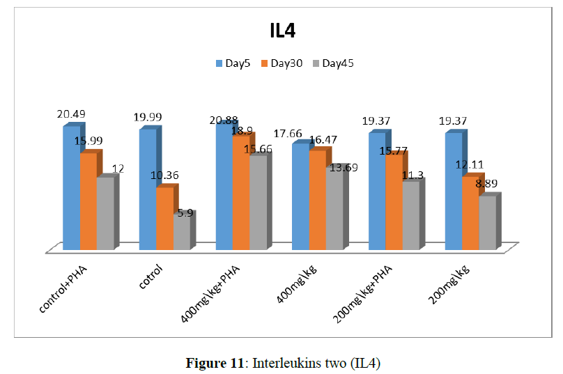
IL-4: group (1) recorded 20.49, which group (2) recorded 19.99. And group (3) 20.88. However, group 4, 5 and 6 recorded 15.9, 17.99 and 17.99, respectively.
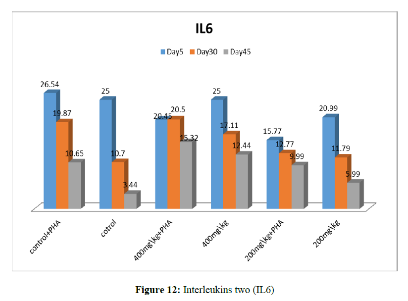
IL-6: Group 1, 2, 3, 4, 5 and 6 recorded, 26.54, 25, 16.32, 25.72, 15.77, and 18.92 respectively.
IL-10: Group 1, 2, 3, 4, 5 and 6 recorded, 30.45, 25.96, 29.60, 50.32, 29.12 and 23.87 respectively.
INFγ: Group 1, 2, 3, 4, 5 and 6 recorded, 19, 20.98, 22.63, 24.32, 19.8 and 25.36 respectively.
Result of 30 days
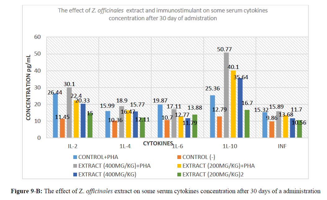
IL-2: the result of group (1) which received PHA at 0.5 I/M showed 26.44. While the control normal saline. Group (2) recorded 11.45. The group (3) which received 400 mg of the extract + the PHA showed 30.1. While the group (4) which received the extract at 400mg only recorded 22.4. However, group (5) received 200 mg extract + PHA recorded 20.33 and group (6) which received the extract only at 200 mg recorded 15.00. Showed in table no (4-B) and figure no (9-B).
IL-4: group (1) recorded 15.99, which group (2) recorded 10.36. And group (3),18.9 However group 4, 5 and 6 recorded 16.47,15.77 and 12.11, respectively.
IL-6: Group 1, 2,3,4,5 and 6 recorded, 19.87, 10.7, 17.11, 12.77,11.79 and 13.88 respectively.
IL-10: Group 1, 2,3,4,5 and 6 recorded, 25.36, 12.79, 50.77, 40.1, 35.64 and 16.7, respectively.
INFγ: Group 1, 2,3,4,5 and 6 recorded 15.32, 9.86, 15.89, 13.68, 11.7 and 10.56, respectively.
Result of 45 days
IL-2: the result of group (1) which received PHA at 0.5 I/M showed 19.56 while the control Normal saline group (2) recorded 9.35. The group (3) which received 400 mg of the extract + the PHA showed 35.87. While the group (4) which received the extract at 400mg only recorded 24.99 However group (5) received 200 mg extract + PHA recorded 22.7 and group (6) which received the extract only at 200 mg recorded 12.66 Showed in table no (4-C) and figure no (9-C).
IL-4: group (1) recorded 12 which group (2) recorded 5.9. And group (3) 12.66. However, group 4, 5 and 6 recorded 13.69, 11.3 and 8.89, respectively.
IL-6: Group 1, 2,3,4,5 and 6 recorded 10.65, 3.44, 15.32, 12.44, 9.99 and 5.9, respectively.
IL-10: Group 1, 2,3,4,5 and 6 recorded 30, 11.3, 60.7, 55.77, 40.3 and 12.99, respectively.
INFγ: Group 1,2,3,4,5 and 6 recorded7.99, 0.88, 5.22, 3.33, 1.7 and 0.9, respectively
Discussion
The present study investigated the chemical structure for the methanolic extract of Punica granatum cortex constituents which were: [5-Hydroxymethylfuraldehyde, Laxiric Acid, Mannitol-manna sugar, Oleanolic acid, palmitic acid, Ellagic acid, Gallic acid and anthocyanidins]. Similar results were obtained by [21].Who reported, twelve compounds were isolated and identified as [oleanolic acid, ursolic acid, palmitic acid, tricin, catechin, rutin, apigenin, apigenin-7-O-glucoside,2S,3S,4S-tri hydroxyl-pentanoic acid, gallic acid, Ellagic acid, β-sitosterol, and daucosterol].
The assessment of chemical structure in the present study is similar to that described by [22]. They were found to be present in the form of calcium pectate in the lamella [23]. Sitosterol, malonic acid, Asiatic acid and alkanes are present in the composition of pomegranate flower. It was expressed that D-mannitol, ellagic acid and gallic acid were present in its alcoholic extract [24]. It is stated that in the pomegranate juice almost all the amino acids are present; valine and methionine are in a very high concentration [25].
Also, the present study investigated the chemical structure for the methanolic extract of Zingiber officinales rhizome constituents which were: [Zingiber, Alpha-Aromadendrene, Borneol, β-Sesquiphellandrene, Gingerols and Shogaol]. Similar results were obtained by [26]. Ginger contains up to three percent of a fragrant essential oil whose main constituents are sesquiterpenoids, with (-) zingiberene as the main component. Smaller amounts of other sesquiterpenoids (β- sesquiphellandrene, bisabolene and farnesene) and a small monoterpenoid fraction (β-phelladrene, cineol, and citral) have also been identified. However, DL McKay and Blumberg mentioned that the characteristic odor and flavor of ginger is caused by a mixture of [Zingerone, Shogaols and Gingerols], volatile oils that compose one to three percent of the weight of fresh ginger [27].
This variation in the chemical constituents although they used methanolic extracts might be attributed to the sensitivity of instruments and methods of analysis such as silica gel column chromatography more sensitive.
In this work, we characterized conjugated linolenic acids (e.g., punicic acid) as the major components of the hydrophilic fraction (80% aqueous methanol extract) from pomegranate (Punica granatum L.) seed oil (PSO) and evaluated their anti-inflammatory potential on some human colon (HT29 and HCT116), liver (HepG2 and Huh7), breast (MCF-7 and MDA-MB-231) and prostate (DU145) cancer lines. Our results demonstrated that punicic acid and its congeners induce a significant decrease of cell viability for two breast cell lines with a related increase of the cell cycle G0/G1 phase respect to untreated cells. Moreover, the evaluation of a great panel of cytokines expressed by MCF-7 and MDA-MB-231 cells showed that the levels of VEGF and nine pro-inflammatory cytokines (IL-2, IL-6, IL-12, IL-17, IP-10, MIP-1α, MIP-1β, MCP-1 and TNF-α) decreased in a dose dependent way with increasing amounts of the hydrophilic extracts of PSO, supporting the evidence of an anti-inflammatory effect. Taken together, the data herein suggest a potential synergistic cytotoxic anti-inflammatory and anti-oxidant role of the polar compounds from PSO [28].
Zingiber officinales rhizome
We found that the mixed formula treatment significantly enhanced IL-10 but decreased IFN-γ and IL-5 production by PHA-stimulated mononuclear cells. The IL-5 production was also decreased by PHA-stimulated lymphocyte. In addition, the COX-2 mRNA expression in stimulated PMN was significantly suppressed after treatment. These results suggest that the new mixed formula treatment is beneficial to the patients with perennial allergic rhinitis via modulating the function of lymphocytes and neutrophils [29].
Disagree Tan and Vanitha mentioned that, ethanol-soluble extracts from the rhizomes of Z. officinale were tested for their action on cytokines and found to promote the secretion of IL-1 and IL-6 in a time- and dose-dependent manner (Hori, et al. 2003). Early isolation experiments showed the presence of highly cytotoxic compounds (diacetylafzelin, diferuloyl methane, feruloyl-pcoumaroyl methane and di-coumaroyl methane) from the species of the family Zingiberaceae [30].
The effect of ginger rhizome powder on hormonal immunity. Results showed that at day (35) of age, ginger rhizome powder in (10 g/kg) increased the HI titer compared to control and 5 g/kg diets. There were not statistically significant differences between treatments for HI titer at (9 and 23) days of age, there were no significant difference between different levels at (9) days of age. but at days (23) of age there was a significant difference between (5 g/kg and 10 g/kg), in conclusion adding ginger rhizome powder in (10 g/kg) improved homural immunity of broilers at 35 days of age. [31].
Ethanol-soluble extracts from the rhizomes of Z. officinales were tested for their action on cytokines and found to promote the secretion of IL-1 and IL-6 in a time- and dose-dependent manner (Hori, et al.2003).Early isolation experiments showed the presence of highly cytotoxiccompounds(diacetylafzelin,diferuloylmethane,feruloyl-p-coumaroylmethane and di-p-coumaroylmethane) from the species of the family Zingiberaceae [32] .
Omar, et al. (2014), was investigated the effects of pomegranate peel extract (PPE) on the outcome of Coccidiosis caused by Eimeria papillata in mice. The data showed that increased mRNA levels of Inducible Nitric Oxide Synthase (INOS), Bcl-2 gene, and of the cytokines interferon gamma (IFN-γ), tumour necrosis factor-α (TNF-α), and interleukin-1β (IL-1β), and as down regulation of mucin gene MUC2 mRNA.
Acknowledgements
The authors would like to express their gratitude's to the ministry of education and the deanship of scientific research - Najran University - Kingdom of Saudi Arabia for their financial and technical support.
References
- Alluwaimi Ahemd, Yehia Hussein and Mohammes Alkhames. Sci. J. King Faisal Univ. 2000, 8(2): p. 1228 73-89.
- Amit Parashar and Shailendra Badal. Int. J. Adv. Biochem. 2018, 2(2): p. 08-12
- Aviram M and Dornfeld L. J Atherosclerosis 2001, 158: p. 195-198.
- Barakat E, Abu-Irmaileh A and Fatma U. Afif . J Ethnopharmacol. 2003, 89: p. 193–197.
- Chidambara MKN, Jayaprakasha GK and Singh RP. J Agric Food Chem. 2002, 50: p. 4791-4795.
- Choudhury D, Amlan, Bhattacharya et al., Food Chem Toxicol. 2010, 48 (10): p. 2872–2880.
- Costantini S, Rusolo F, Devito V et al., J Molecules. 2014, 19(6): p. 8644-8660.
- Darrell R Boverhof, Greg Ladics, Bob Luebke et al., Regul Toxicol Pharmacol. 2014, 68: p. 96-107.
- Deniz Azhir, Afshin Zakeri and Ali et al., Euro J Exp Bio. 2012, 2(6): p. 2090-2092.
- McKay DL and Blumberg JB. Phytother Res. 2006, 20(7): p. 519-530.19(6): p. 8644-8660.
- Gardiner P and KJ Kemper. Pediatr Rev. 2000. 21(2): p. 44–57
- Hassan AA, Abdel-Ati KE and Shehata AS. Egypt J Plant Breeding. 2014, 18(4): p. 597-606.
- Hori Y, Miura T, Hirai Y et al., 62: p. 613-617.
- Huang, Tom H, Pharm WB et al., J. Cardiovasc. Pharmacol. 2005, 46(6): p. 856-862.
- Jhon J Rojas, Veronica J Ochoa, Saul A Ocampo et al., BMC Complement Altern Med. 2006, 6: p. 2.
- Kim JS, Sa Im, Park Hye et al., Arch Pharm Res. 2008, 31(4): p. 415–418.
- Meltem Türkyılmaz, Şeref Tağı, Ufuk Dereli et al., J Food Chem. 2013, 138(2): p. 1810-1818.
- Nicholas and Trulove F. ProQuest Dissertations Publishing, 2013, p. 1539500.
- Nievergelt A, Huonker P, Schoop R et al., J Bioorg Med Chem. 2010, 18(9): p. 3345-51.
- Oliver Bever BEP. Medicinal plants in tropical West Africa. 1986.
- Omar SO, Amer Mohamed A, Dkhil Wafaa M et al., 2014.
- Oyagbemi AA, Saba AB and Azeez OI. J Biofactors 2010, 36 (3):169–178.
- Premalatha Balachandran and Rajgopal Govindarajan. Pharmacol. Res. 2002, 5 (1): p. 19-30.
- Rhode J, Fogoros S, Zick S et al., BMC complement med ther. 2007, 7: p.44.
- Sharrif Moghaddasi Mohammad and Hamed Haddad Kashani. J. Med. Plant Res. 2012, 6(40): p. 5306-5310.
- Sien-Hung Yang, Chuang-Ye, Hong and Chia-Li, Yu. Elsevier Science. 2001.
- Snedecor GW and Cocharn WG. Statistical Methods. 1989.
- Srivastava Rashmi, Chauhan Deepa and Chauhan JS. Indian J. Chem. 2001, 2: p. 170-172.
- Sugeng Riyanto. Indo J chem. 2007, 7(1): p. 93-96.
- Tan, Benny KH and Vanitha J. J Bentham Science Publishers. 2004, 11: p. 1423-1430.
- Vladimir, Badmaev and Maja and Nowakowski. Phytother Res. 2010, 14: p. 245–249
- Zhong and Yao Cai. Pub Med. 2004, 37(5): p. 804-7.

